Retinoblastoma genetics centers on one tumor suppressor gene, RB1, on chromosome 13q14.
Every affected child needs genetic assessment, because the result shapes care for the whole family.
The Two-Hit Model in Retinoblastoma Genetics
Knudson proposed in 1971 that retinoblastoma requires two mutational events, and today we know both RB1 alleles in a developing retinal cell must lose function. When the first hit is in the germline, every retinal cell already carries it. Therefore, one further hit anywhere can start a tumor, which explains bilateral and multifocal disease.
Germline vs Somatic RB1
A germline variant is present in blood and in nearly all body cells. In contrast, a somatic variant exists only within the tumor. Nonheritable disease arises when both hits occur in one retinal cell after conception, so the child cannot pass it on.
Fundus Explorer Pro
Photograph the retinal findings described here with the phone already in your pocket — 22 D optics and built-in illumination in one handheld unit.
From Choroida — the team behind this siteHeritable Disease and Family History
About 40% of children have heritable disease, while about 60% have the nonheritable form. However, most germline variants arise de novo, and as a result, the parents are usually unaffected and family history is often negative.
Laterality in Retinoblastoma Genetics
Bilateral disease almost always reflects a germline or mosaic RB1 variant. Most unilateral cases are somatic, but a minority carry a germline variant, so laterality alone cannot exclude heritable risk. Early onset or several tumors in one eye should also raise suspicion.
Who Needs Testing
- Every child with retinoblastoma, whether unilateral or bilateral
- Parents of an affected child, once the child’s variant is known
- Siblings and offspring of any confirmed carrier
- Adult survivors treated before testing became available
- Children with a known 13q deletion, who also need retinal examination
Blood and Tumor Testing in Retinoblastoma Genetics
Testing usually starts with a blood sample from the affected child, and when an enucleated eye is available, tumor DNA adds valuable information. The laboratory first identifies both tumor hits, then searches the blood for those exact variants. If neither appears in blood, heritable risk falls considerably, but low-level mosaicism can escape detection, so counseling should mention residual risk.
Mosaicism in Retinoblastoma Genetics
A de novo variant may arise after fertilization, leaving only some cells affected, and mosaic children can still develop bilateral disease. Their offspring face a transmission risk below 50%, but the risk remains real. Similarly, an unaffected parent may carry low-level mosaicism, so siblings still need examination.
Low-Penetrance Variants
Some RB1 variants retain partial protein function, including certain missense, splice and promoter changes. Carriers may develop unilateral tumors, retinoma, or no tumor at all, and consequently these families may show skipped generations that mimic sporadic disease.
MYCN-Amplified Tumors
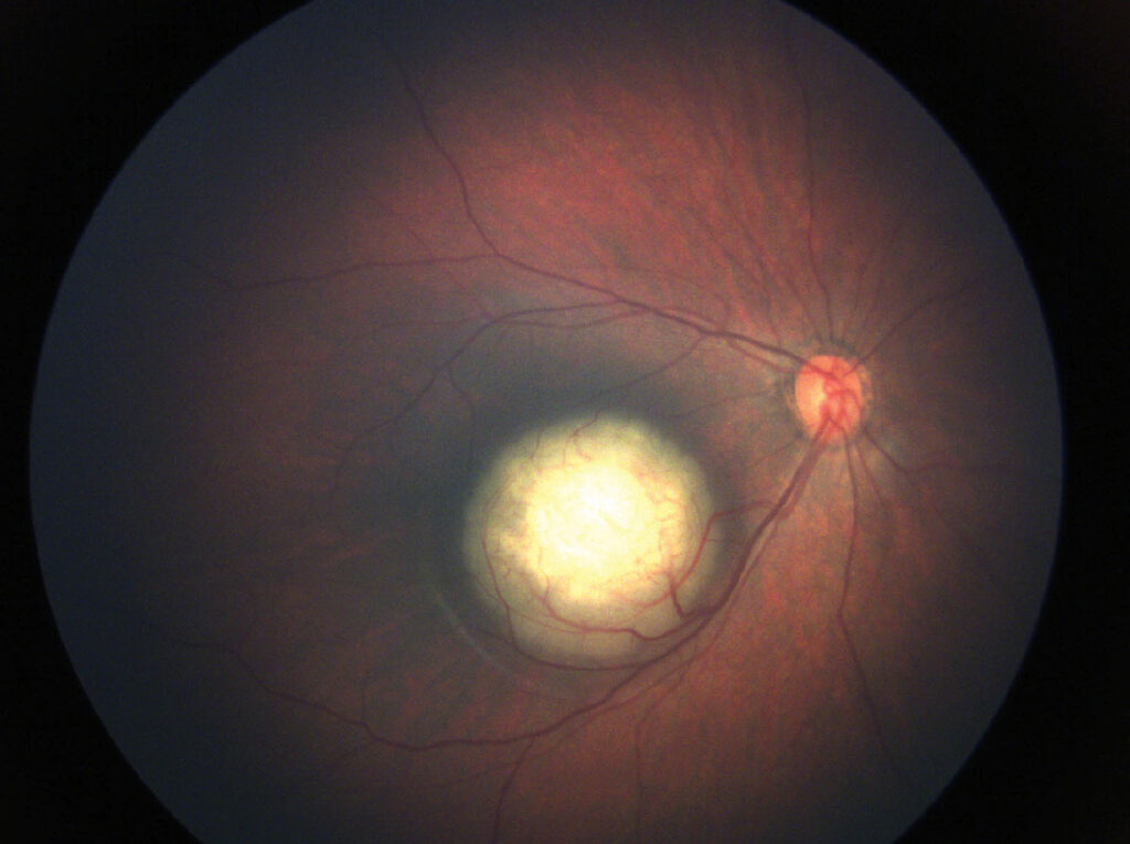
A small group of unilateral tumors has normal RB1 alleles and MYCN amplification instead. These tumors typically present in very young infants and often look large at diagnosis. Current data suggest little germline risk in this subgroup, although numbers remain limited.
13q Deletion Syndrome
Large 13q14 deletions remove RB1 together with neighboring genes, and affected children may show developmental delay and dysmorphic features. Therefore, retinoblastoma plus these findings calls for chromosomal microarray testing.
Sibling Screening in Retinoblastoma Genetics
Siblings of an affected child need dilated fundus examinations from early infancy, while examination frequency depends on calculated risk and many toddlers need examination under anesthesia. When the familial variant is known, targeted testing shows who truly needs surveillance.
Then a sibling who tests negative can usually stop intensive examinations, while a carrier continues frequent checks, because small tumors respond well to focal therapy (see ICRB grouping).
Offspring of Survivors
A germline carrier passes the variant to each child with a 50% probability. Penetrance for most null variants is high, so most carriers develop tumors. Therefore, early neonatal testing helps plan the screening schedule, and some families also consider prenatal or preimplantation genetic testing with a genetics team.
Second Cancers in Heritable Disease
Germline carriers face a lifelong excess risk of new primary cancers, with osteosarcoma and soft tissue sarcomas as the classic examples and melanoma also appearing more often. External beam radiation raises sarcoma risk within the treated field, especially in infancy. For this reason, modern protocols favor chemotherapy and focal treatment, and survivors should avoid unnecessary radiation.
Counseling Families on Retinoblastoma Genetics
Parents often ask whether they caused the disease, and for most families the honest answer is no, because the variant arose by chance. Explain risk in plain numbers and repeat the message at later visits, and revisit counseling when survivors reach adolescence and plan their own families. For a broader framework, see genetic testing for inherited retinal disease.
Pitfalls to Avoid
- Labeling unilateral disease nonheritable without testing
- Ignoring mosaicism when parental blood is negative
- Stopping sibling screening before genetic results return
- Forgetting to counsel adult survivors about their children


Document what you see
Two smartphone imaging tools built for everyday clinic use — one for the slit lamp, one for the fundus.
From Choroida — the team behind this siteReferences
- Knudson AG Jr. Mutation and cancer: statistical study of retinoblastoma. Proc Natl Acad Sci U S A. 1971;68:820-823.
- Dimaras H, Kimani K, Dimba EA, et al. Retinoblastoma. Lancet. 2012;379:1436-1446.
- Rushlow DE, Mol BM, Kennett JY, et al. Characterisation of retinoblastomas without RB1 mutations: genomic, gene expression, and clinical studies. Lancet Oncol. 2013;14:327-334.
- Skalet AH, Gombos DS, Gallie BL, et al. Screening children at risk for retinoblastoma: consensus report from the American Association of Ophthalmic Oncologists and Pathologists. Ophthalmology. 2018;125:453-458.
- Kleinerman RA, Tucker MA, Tarone RE, et al. Risk of new cancers after radiotherapy in long-term survivors of retinoblastoma: an extended follow-up. J Clin Oncol. 2005;23:2272-2279.
Test yourself
A few questions straight from this article.
-
Under the two-hit model of retinoblastoma, why does a germline RB1 first hit explain bilateral and multifocal tumors?
Both RB1 alleles must lose function; with a germline first hit every retinal cell already carries it, so one further hit anywhere can start a tumor. -
About what proportion of children with retinoblastoma have heritable disease?
About 40% of children have heritable retinoblastoma and about 60% have the nonheritable form. -
Why is family history often negative in heritable retinoblastoma?
Most germline RB1 variants arise de novo, so the parents are usually unaffected and family history is often negative. -
Which children with retinoblastoma need RB1 genetic testing?
Bilateral disease almost always reflects a germline or mosaic variant, but some unilateral cases also carry one, so laterality alone cannot exclude heritable risk. -
When an enucleated retinoblastoma eye is available, how does RB1 testing usually proceed?
The laboratory identifies both tumor hits, then searches blood for those exact variants; absence in blood lowers heritable risk, though low-level mosaicism can escape detection. -
Why may families with low-penetrance RB1 variants show skipped generations that mimic sporadic retinoblastoma?
Some missense, splice and promoter variants retain partial RB1 function, so carriers may develop unilateral tumors, retinoma or no tumor at all. -
Which features characterize MYCN-amplified retinoblastoma?
A small group of unilateral tumors has normal RB1 alleles and MYCN amplification, presents in very young infants, often looks large, and appears to carry little germline risk. -
A child with retinoblastoma also has developmental delay and dysmorphic features. Which test is indicated?
Large 13q14 deletions remove RB1 with neighboring genes, causing developmental delay and dysmorphic features, so this combination calls for chromosomal microarray testing. -
Why do modern protocols for heritable retinoblastoma favor chemotherapy and focal treatment over external beam radiation?
Germline carriers face lifelong excess risk of osteosarcoma, soft tissue sarcomas and melanoma, and external beam radiation raises sarcoma risk in the field, especially in infancy. -
When a familial RB1 variant is known, what happens to a sibling who tests negative?
Targeted testing shows who truly needs surveillance: a negative sibling can usually stop intensive examinations, while a carrier continues frequent checks.

Writes and reviews the clinical articles on Educate. Every post is checked against peer-reviewed sources before it goes live.