
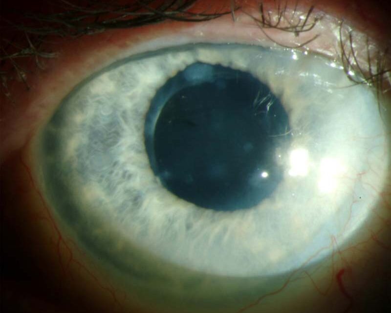
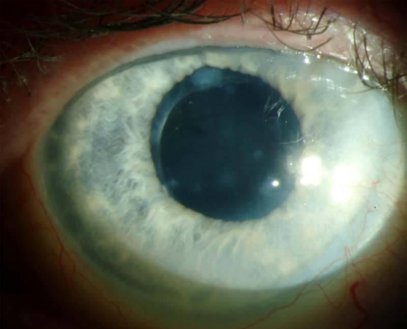
Salzmann nodular degeneration produces elevated, bluish-white to gray subepithelial nodules on the cornea, typically arising after years of chronic ocular surface inflammation, and despite its appearance and the fibrotic tissue that composes it, it is a degenerative rather than an inflammatory or infectious process by the time it presents, which changes the management away from anti-inflammatory or antimicrobial treatment and toward mechanical removal when the nodules cause enough trouble.
What the Nodules Are
The nodules consist of subepithelial fibrous tissue, deposited in a zone just beneath the epithelium and above Bowman layer, thought to result from a long-standing reparative response to chronic epithelial irritation and inflammation, even though by the time the nodules are clinically apparent, active inflammation may have largely subsided.
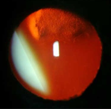
They are typically multiple, arranged in an annular or scattered pattern, most often located in the mid-peripheral cornea, though they can occur centrally and affect the visual axis directly.
Associated Conditions
Salzmann nodular degeneration is frequently preceded by, or occurs in association with, chronic ocular surface disease.
- Chronic blepharitis and meibomian gland dysfunction
- Phlyctenular keratoconjunctivitis
- Vernal and atopic keratoconjunctivitis
- Trachoma, in endemic regions
- Prior corneal surgery or trauma
- Dry eye disease of various causes
- In some patients, no clear preceding condition is identified
All-fit Slit-Lamp Adapter
Record and share exactly what you see at the slit lamp. One adapter fits any slit lamp or surgical microscope — and any smartphone.
From Choroida — the team behind this siteClinical Features
- Elevated, bluish-white to grayish, well-circumscribed nodules, typically in the mid-periphery, sometimes centrally
- Foreign body sensation, particularly when a nodule has an irregular, elevated surface that disrupts normal lid closure and blinking
- Reduced vision from irregular astigmatism when nodules are near or within the visual axis, or from tear film disruption over the irregular surface
- A generally slow, progressive course over years
Evaluation
Diagnosis is usually clinical, based on the characteristic slit-lamp appearance.
- Corneal topography helps quantify the irregular astigmatism contributing to visual symptoms and provides a baseline for comparison after treatment
- Assessment for underlying or coexisting ocular surface disease, since untreated chronic surface inflammation increases the risk of nodule recurrence after removal
- Anterior segment OCT can help characterize nodule depth, useful for surgical planning
Management
Addressing the Ocular Surface
Treating any underlying blepharitis, meibomian gland dysfunction, or dry eye disease is an important first step, both for symptomatic improvement and to reduce the risk of recurrence after any surgical treatment of the nodules themselves.
Observation
Small, peripheral, minimally symptomatic nodules that do not significantly affect vision or comfort can be observed without intervention.
Superficial Keratectomy
Manual superficial keratectomy, removing the nodules and the affected superficial stroma, is the primary treatment for visually significant or symptomatic nodules, generally straightforward given the relatively superficial plane of the disease.
Phototherapeutic Keratectomy
Excimer laser PTK can be used as an adjunct or alternative to manual keratectomy, smoothing the corneal surface after nodule removal and potentially reducing irregular astigmatism more precisely than manual technique alone.
Recurrence
Nodules can recur after removal, particularly when underlying chronic ocular surface disease is not adequately controlled, so ongoing management of blepharitis, dry eye, or other contributing conditions continues after surgical treatment, not just before it.
Prognosis
Surgical removal generally provides good symptomatic relief and often meaningful visual improvement when nodules were affecting the visual axis or causing significant irregular astigmatism, though a minority of patients experience recurrence over subsequent years, particularly if the predisposing ocular surface condition persists.


Document what you see
Two smartphone imaging tools built for everyday clinic use — one for the slit lamp, one for the fundus.
From Choroida — the team behind this siteReferences
- Farjo AA, Halperin GI, Syed N, Sutphin JE, Wagoner MD. Salzmann’s nodular corneal degeneration clinical characteristics and surgical outcomes. Cornea. 2006;25:11-15.
- Das S, Link B, Seitz B. Salzmann’s nodular degeneration of the cornea: a review and case series. Cornea. 2005;24:772-777.
- Graue-Hernandez EO, Mannis MJ, Eliasieh K, Greasby TA, Beckett LA, Bradley JC. Salzmann nodular degeneration. Cornea. 2010;29:283-289.
- Maharana PK, Sharma N, Das S, et al. Salzmann’s nodular degeneration. Ocul Surf. 2016;14:20-30.
Test yourself
A few questions straight from this article.
-
Salzmann nodules consist of fibrous tissue deposited in which corneal plane?
The nodules are subepithelial fibrous tissue deposited in the zone just beneath the epithelium and above Bowman layer, which is why the disease is surgically superficial. -
Why is Salzmann nodular degeneration not managed with anti-inflammatory or antimicrobial therapy?
Although it follows years of surface inflammation, by presentation the process is degenerative rather than inflammatory or infectious, so management shifts toward mechanical removal. -
Where are Salzmann nodules most often located on the cornea?
Nodules are typically multiple and arranged in an annular or scattered pattern, most often in the mid-periphery, though they can occur centrally and involve the visual axis. -
Which ocular surface condition is associated with Salzmann nodular degeneration?
Associated chronic surface disease includes blepharitis and meibomian gland dysfunction, phlyctenular keratoconjunctivitis, vernal and atopic disease, trachoma, prior surgery and dry eye. -
Foreign body sensation in Salzmann nodular degeneration arises from which mechanism?
Discomfort is particularly reported when a nodule has an irregular, elevated surface that disrupts normal lid closure and blinking. -
Reduced vision in Salzmann nodular degeneration results from which mechanisms?
Vision falls from irregular astigmatism when nodules sit near or within the visual axis, and from disruption of the tear film over the irregular surface. -
What is the typical natural course of Salzmann nodular degeneration?
The course is generally slow and progressive over years, which is one reason small, minimally symptomatic nodules can simply be observed. -
What is anterior segment OCT used for in Salzmann nodular degeneration?
Anterior segment OCT helps characterize how deep the nodules extend, which is useful when planning surgical removal. -
What is the primary treatment for visually significant or symptomatic Salzmann nodules?
Manual superficial keratectomy removes the nodules and affected superficial stroma and is the primary treatment, generally straightforward given the superficial plane of disease. -
What most increases the risk of Salzmann nodule recurrence after removal?
Nodules recur particularly when underlying chronic surface disease is not controlled, so treatment of blepharitis and dry eye continues after surgery as well as before it.
