
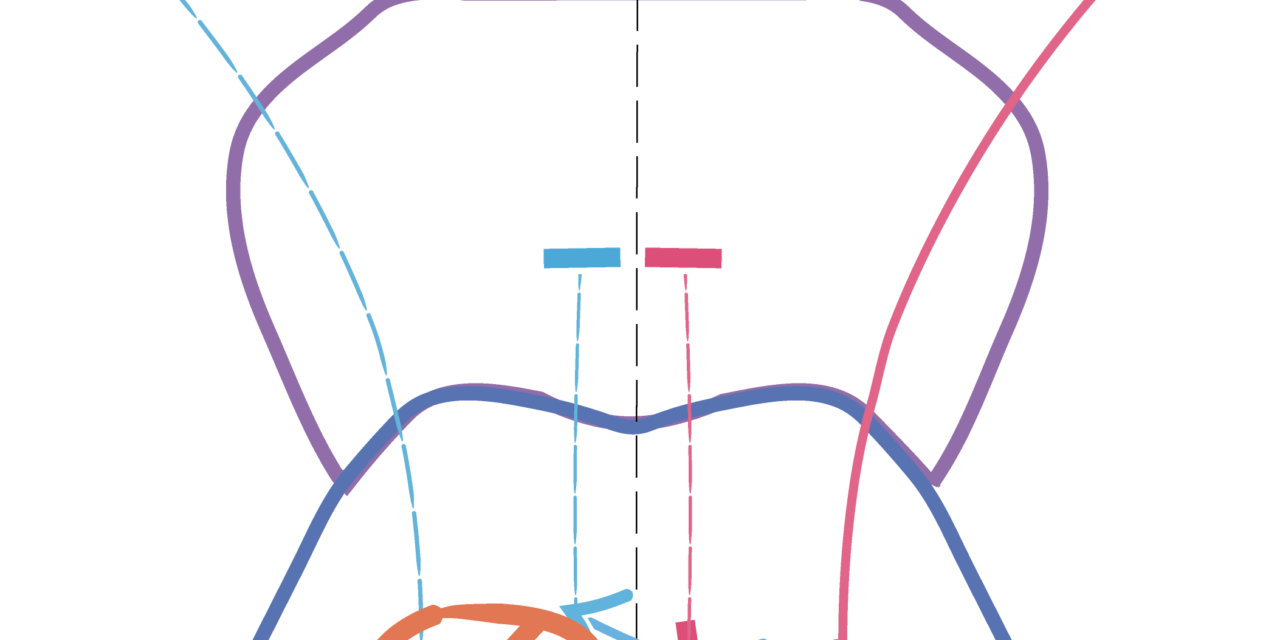
One-and-a-half syndrome is a distinctive eye movement disorder that lets a clinician localize a lesion to a specific, small area of the dorsal pons from the bedside examination alone, before imaging even confirms it.
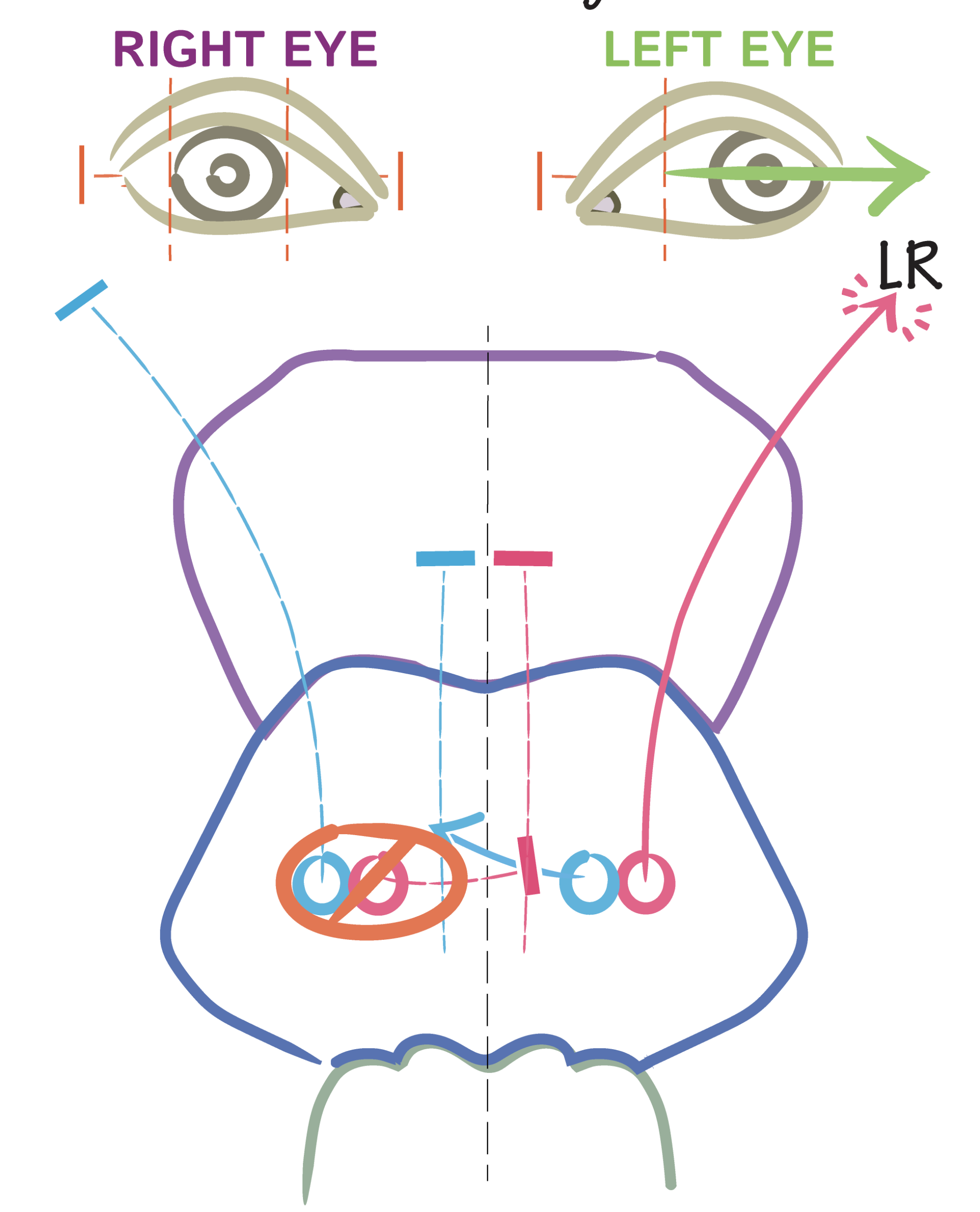
The name describes the deficit precisely: a complete horizontal gaze palsy to one side (the “one”) plus an internuclear ophthalmoplegia on attempted gaze to the other side (the “half”), leaving only a single functioning eye movement, abduction of the eye opposite the gaze palsy.
The Anatomy Behind the Name
The syndrome results from a single lesion that damages both the paramedian pontine reticular formation (or the abducens nucleus itself) on one side, and the adjacent medial longitudinal fasciculus on the same side.
The pontine gaze center lesion eliminates all horizontal gaze, both saccadic and pursuit, toward that side, while the medial longitudinal fasciculus lesion, which is responsible for internuclear communication that drives the adducting eye during a conjugate horizontal saccade, eliminates adduction of the ipsilateral eye during attempted gaze to the opposite side.
The only horizontal movement left is abduction of the eye contralateral to the lesion, when the patient looks away from the side of the lesion.
Clinical Findings
- No horizontal eye movement of any kind toward the side of the lesion, in either eye
- On attempted gaze away from the lesion, the ipsilateral eye fails to adduct (the internuclear ophthalmoplegia component) while the contralateral eye abducts, often with nystagmus
- Vertical eye movements and convergence are typically preserved, since these pathways are largely separate from the horizontal gaze system
- A subtle exotropia of the eye ipsilateral to the lesion is sometimes seen at rest, called paralytic pontine exotropia, from the unopposed action of the intact contralateral medial rectus pathway
- Facial nerve involvement is common, since the seventh nerve fascicle wraps around the sixth nerve nucleus in the same region, and a facial palsy on the side of the lesion is a useful supporting clue
Causes
- Multiple sclerosis, a common cause, particularly in younger patients, from a demyelinating plaque in the dorsal pons
- Brainstem stroke, particularly in older patients with vascular risk factors, from infarction in the territory of the basilar artery’s paramedian branches
- Pontine tumor
- Brainstem hemorrhage
- Less commonly, infection, trauma, or other structural lesions of the dorsal pons
The likely cause is often suggested by the patient’s age and the tempo of onset: sudden onset in an older patient with vascular risk factors suggests stroke, while a younger patient with a more subacute course and other neurological symptoms suggests demyelinating disease.
Evaluation
- MRI of the brain, focused on the pons, is the key imaging study and typically shows a small, well-localized lesion corresponding to the clinical findings
- Assessment for other neurological signs, including facial weakness, which supports the anatomic localization
- Investigation directed by the suspected cause: vascular risk factor assessment and stroke workup for older patients, or a demyelinating disease workup, including MRI of the rest of the brain and spinal cord and consideration of lumbar puncture, for younger patients or those with a pattern suggesting multiple sclerosis
Management
Treatment addresses the underlying cause rather than the eye finding itself: stroke management and secondary prevention for vascular disease, disease-modifying therapy for multiple sclerosis, or targeted treatment for a tumor or other structural lesion.
The eye movement abnormality itself is managed symptomatically if diplopia or visual discomfort persists once the acute phase has settled, sometimes with prism correction or, rarely, strabismus surgery for a fixed residual deviation.
Prognosis
Recovery of eye movements depends on the underlying cause and the extent of the lesion, ranging from significant improvement over weeks in a demyelinating plaque to a more fixed deficit after an ischemic stroke.
Even with residual gaze limitation, many patients adapt reasonably well functionally, particularly since vertical gaze and convergence are typically spared, preserving much of the visual field accessible through head movement and vertical eye movement.


Document what you see
Two smartphone imaging tools built for everyday clinic use — one for the slit lamp, one for the fundus.
From Choroida — the team behind this siteReferences
- Fisher CM. Some neuro-ophthalmological observations. J Neurol Neurosurg Psychiatry. 1967;30:383-392.
- Wall M, Wray SH. The one-and-a-half syndrome: a unilateral disorder of the pontine tegmentum. Neurology. 1983;33:971-980.
- Eggenberger E. Eight-and-a-half syndrome: one-and-a-half syndrome plus cranial nerve VII palsy. J Neuroophthalmol. 1998;18:114-116.
- Leigh RJ, Zee DS. The Neurology of Eye Movements. 5th ed. New York: Oxford University Press; 2015.
