
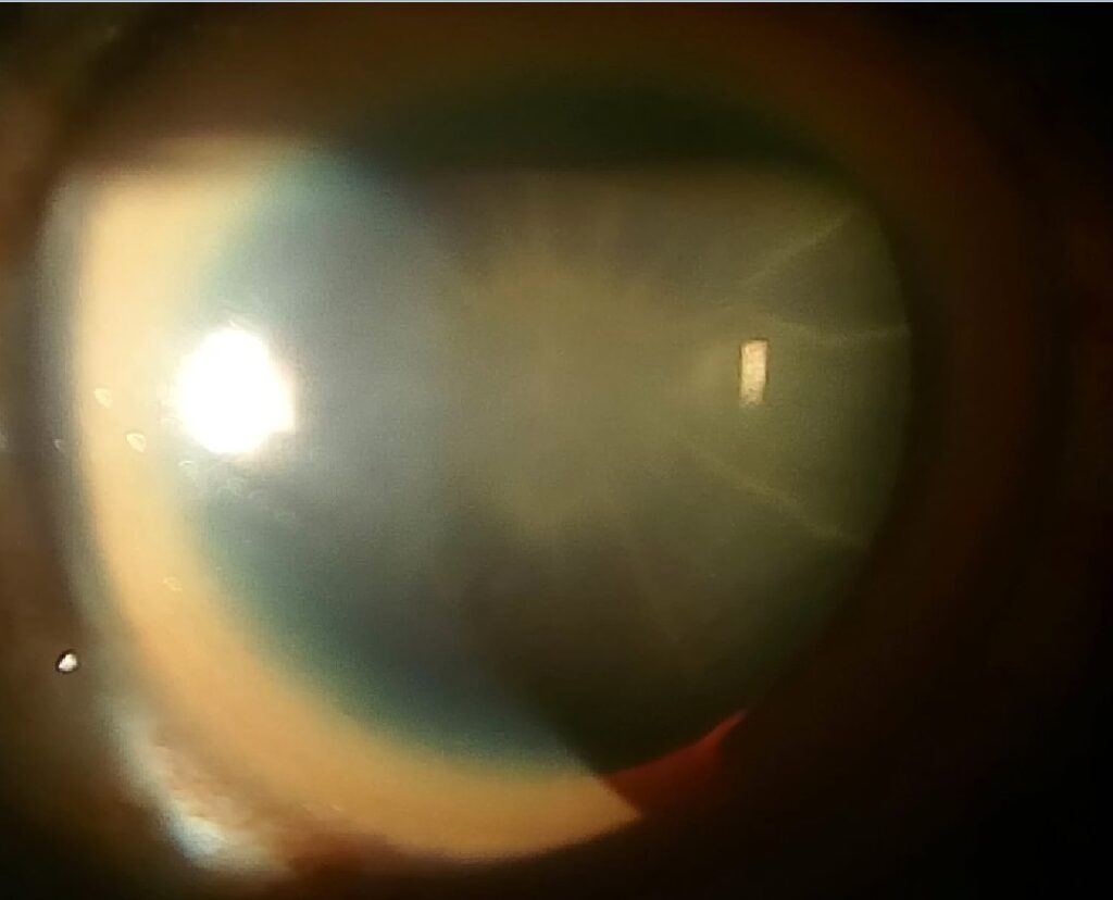
Sunflower cataract is a distinctive pattern of copper deposition on the anterior lens capsule, seen in Wilson’s disease, an autosomal recessive disorder of copper metabolism caused by mutations in the ATP7B gene.
Copper that cannot be properly excreted into bile accumulates in tissues throughout the body, and the eye — specifically Descemet’s membrane and the anterior lens capsule — is one of the sites where this accumulation becomes visible on ordinary slit-lamp examination.
Sunflower cataract is far less commonly discussed than the Kayser-Fleischer ring, its corneal counterpart, but the two findings share the same underlying mechanism and often coexist in the same patient, making a combined slit-lamp exam a useful, low-cost screening step in suspected Wilson’s disease that can support the diagnosis well before biochemical results return.
Pathophysiology
Free copper circulating in excess, not bound to ceruloplasmin, deposits in tissues with high metabolic turnover or fixed structural proteins that bind it readily.
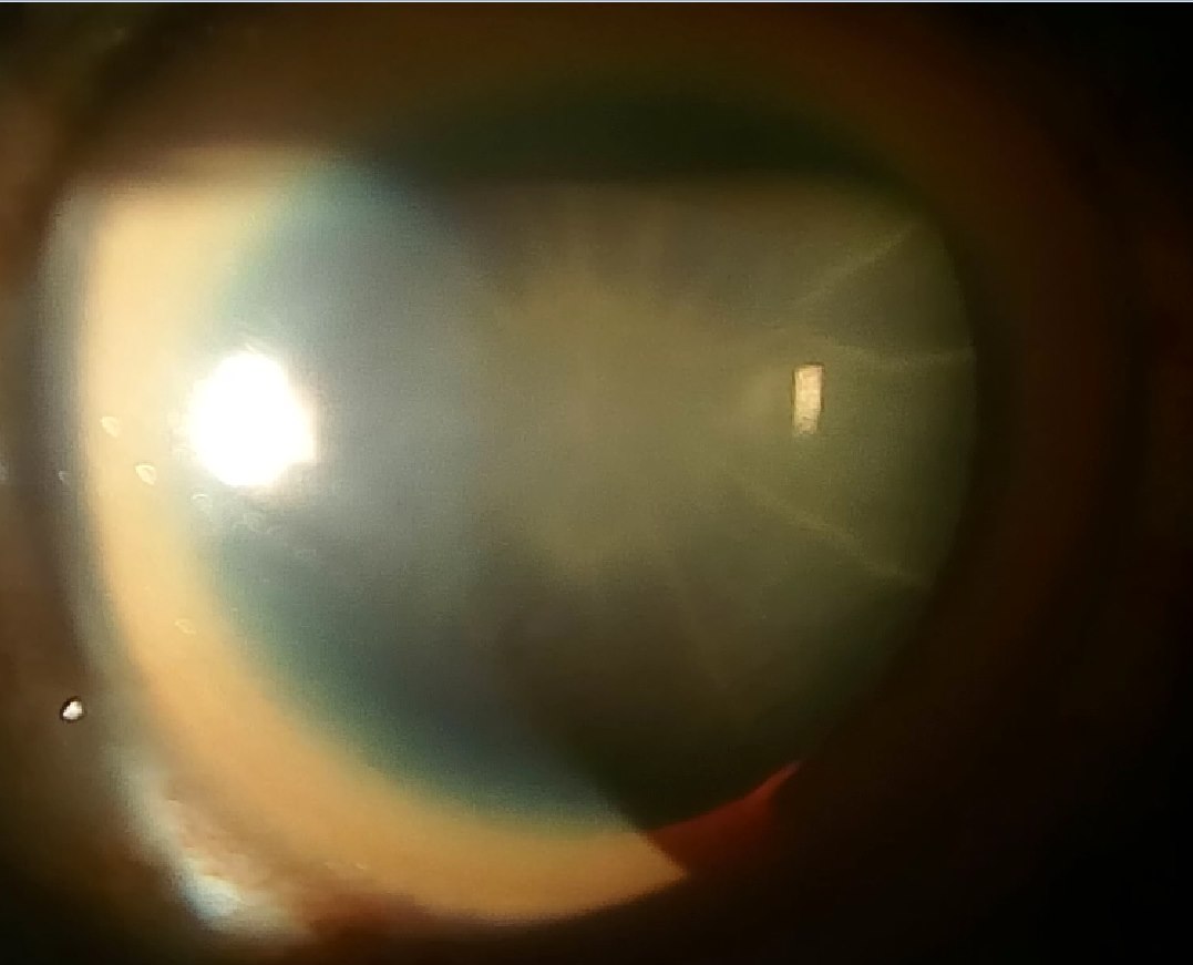
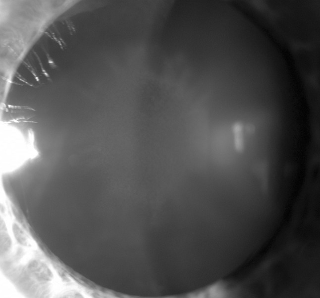
In the eye, this happens at Descemet’s membrane (producing the Kayser-Fleischer ring) and at the anterior lens capsule, where copper accumulates in a radiating, petal-like pattern around the central axis, hence the name, evoking the arrangement of a sunflower’s petals around its disc.
Unlike most cataract types discussed elsewhere on this site, sunflower cataract does not significantly opacify the lens or reduce vision in most patients; it is a pigmentary deposit rather than a true opacifying cataract in the functional sense, which is part of why it is easy to overlook without a deliberate slit-lamp search.
The term “cataract” is, in a sense, a historical misnomer that has simply stuck, since the finding functions clinically as a diagnostic marker of copper overload rather than as a vision-threatening lens opacity requiring its own dedicated ophthalmic management.
Clinical Findings
- A greenish-brown to golden, radially arranged (stellate) pigmentation on the anterior lens capsule, best seen with a dilated pupil and careful slit-lamp technique
- Bilateral, though sometimes asymmetric in degree
- Usually visually insignificant on its own
- Frequently coexists with a Kayser-Fleischer ring, though the two findings can occur independently of each other
Because the finding rarely causes symptoms, it is typically discovered incidentally during a slit-lamp exam performed for another reason, or specifically sought once Wilson’s disease is already suspected on other clinical or laboratory grounds.
All-fit Slit-Lamp Adapter
Record and share exactly what you see at the slit lamp. One adapter fits any slit lamp or surgical microscope — and any smartphone.
From Choroida — the team behind this siteRelationship to Kayser-Fleischer Ring
The Kayser-Fleischer ring — copper deposition in Descemet’s membrane at the corneal periphery, appearing as a golden-brown to greenish ring best seen at the superior and inferior limbus first — is the more widely known and more sensitive ocular sign of Wilson’s disease, present in a large majority of patients with neurologic involvement and a smaller proportion of those with liver-predominant disease.
Sunflower cataract is less consistently present and is generally a secondary, confirmatory finding rather than a primary screening tool on its own, though when both signs are present together, the combination is highly specific for chronic copper overload.
However, neither finding alone, nor both together, is fully sensitive, meaning their absence does not rule out Wilson’s disease, especially early in the disease course.
This limited sensitivity is an important point to communicate when counseling a patient or family anxious about a normal slit-lamp exam early in a workup that remains otherwise clinically concerning for the diagnosis.
Diagnostic Context
Wilson’s disease should be considered in any patient, a child, adolescent, or young adult, presenting with unexplained liver disease, a movement disorder, psychiatric symptoms, or a combination of these, especially with a family history of any of them.
Serum ceruloplasmin (typically low), 24-hour urinary copper excretion (typically elevated), and, when needed, liver biopsy with quantitative copper measurement or genetic testing for ATP7B mutations establish the diagnosis alongside the ocular findings, with the overall diagnostic picture typically built from a combination of several of these tests rather than any single result alone.
A dedicated slit-lamp exam for both the Kayser-Fleischer ring and sunflower cataract should be requested in any patient undergoing workup for suspected Wilson’s disease, since these findings support the diagnosis rapidly and non-invasively while other testing is pending, though a negative exam does not exclude the diagnosis and should not delay further workup.
Management
Treatment of Wilson’s disease itself — copper chelation with D-penicillamine or trientine, or zinc therapy to reduce intestinal copper absorption — addresses the underlying disease and is typically followed by gradual regression of both the Kayser-Fleischer ring and the sunflower cataract over months to a few years, providing a useful, if slow, marker of treatment response.
The sunflower cataract itself does not require any ocular-specific treatment, since it does not threaten vision in the way that other cataract types can, and standard cataract surgery decisions, if ever needed later in life for an unrelated, genuine age-related cataract, are made independently of this copper-related pigmentary finding.
Regular follow-up slit-lamp exams can be a helpful adjunct to systemic markers (ceruloplasmin, urinary copper) in tracking response to chelation or zinc therapy over treatment, because the ocular signs reflect the same underlying copper burden being addressed systemically.
Because regression of the ocular findings lags behind biochemical improvement, a persistently visible sunflower cataract or Kayser-Fleischer ring early in treatment should not be interpreted as treatment failure, provided the systemic copper markers are trending appropriately in the expected direction.


Document what you see
Two smartphone imaging tools built for everyday clinic use — one for the slit lamp, one for the fundus.
From Choroida — the team behind this siteReferences
- Roberts EA, Schilsky ML. Diagnosis and treatment of Wilson disease: an update. Hepatology.
- European Association for the Study of the Liver. EASL clinical practice guidelines: Wilson’s disease. Journal of Hepatology.
- Walshe JM. The eye in Wilson disease. QJM.
- American Academy of Ophthalmology. Basic and Clinical Science Course, Section 11: Lens and Cataract.
Test yourself
A few questions straight from this article.
-
In Wilson's disease, sunflower cataract represents copper deposition at which ocular site?
Copper accumulates in a radiating, petal-like pattern on the anterior lens capsule, producing the sunflower appearance. -
Mutations in which gene cause Wilson's disease, the disorder underlying sunflower cataract?
Wilson's disease is an autosomal recessive disorder of copper metabolism caused by mutations in the ATP7B gene. -
In Wilson's disease, copper accumulates in tissues because it cannot be properly excreted into which route?
Copper that cannot be excreted into bile builds up in tissues throughout the body, including the cornea and lens capsule. -
What is the classic slit-lamp appearance of sunflower cataract in Wilson's disease?
The deposit is a greenish-brown to golden, radially arranged pigmentation on the anterior capsule, best seen through a dilated pupil. -
The Kayser-Fleischer ring of Wilson's disease reflects copper deposition in which corneal layer?
The ring is copper deposited in Descemet's membrane at the corneal periphery, seen first at the superior and inferior limbus. -
Which ocular sign is the more sensitive and more widely recognised marker of Wilson's disease?
The Kayser-Fleischer ring is the more sensitive sign; sunflower cataract is less consistently present and serves as a confirmatory finding. -
A young adult with unexplained liver disease has a normal slit-lamp exam. What does this indicate about Wilson's disease?
Neither ocular sign is fully sensitive, so their absence does not rule out Wilson's disease, especially early in the disease course. -
Which laboratory pattern typically supports a diagnosis of Wilson's disease?
Serum ceruloplasmin is typically low and 24-hour urinary copper excretion typically elevated, alongside the ocular findings. -
Which agent treats Wilson's disease by reducing intestinal copper absorption rather than chelating copper?
Chelation uses D-penicillamine or trientine, whereas zinc therapy works by reducing copper absorption from the intestine. -
A patient on chelation still shows a sunflower cataract while copper markers improve. How should this be interpreted?
Regression of the ocular signs lags behind biochemical improvement and takes months to a few years, so persistence is not treatment failure.

Writes and reviews the clinical articles on Educate. Every post is checked against peer-reviewed sources before it goes live.


