
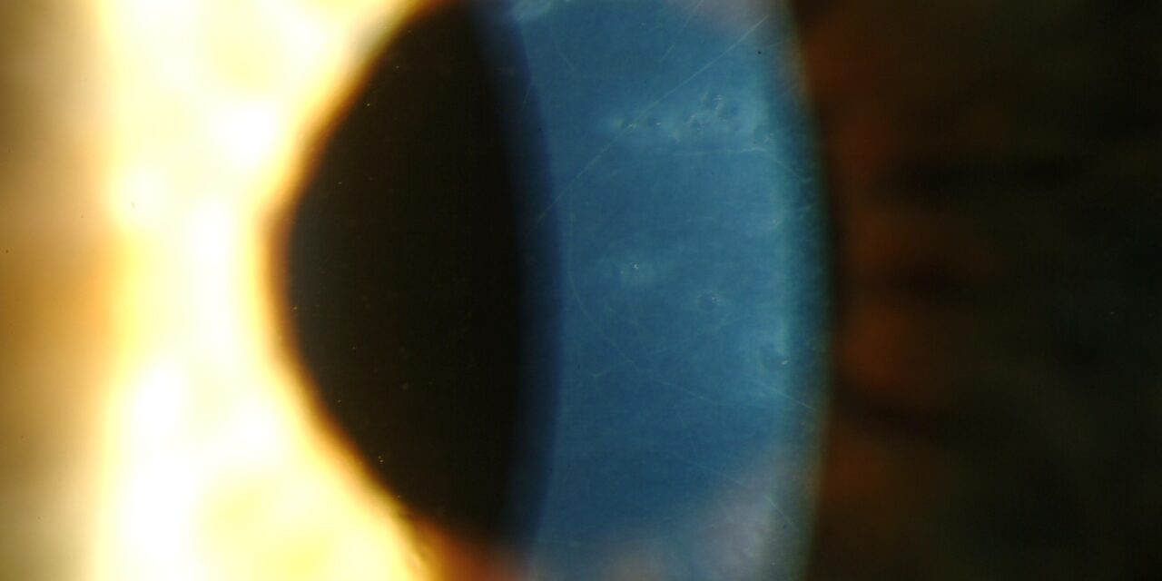
Posterior polymorphous corneal dystrophy (PPCD) is a bilateral, autosomal dominant endothelial dystrophy notable for how variable its clinical impact is: many affected individuals carry the diagnosis for life without ever developing significant visual symptoms, discovered only incidentally on a slit-lamp exam performed for another reason, while a smaller subset develop progressive corneal edema or glaucoma requiring active treatment.
This wide range of severity, more than almost any other single feature, is what shapes how the condition is actually managed in practice, since the same genetic diagnosis can mean lifelong observation for one patient and active treatment for another.
What Makes PPCD Different From Other Endothelial Dystrophies
Unlike Fuchs endothelial corneal dystrophy, discussed in its own dedicated article on this site, where the endothelium progressively loses cells and function over years, the corneal endothelial cells in PPCD undergo an unusual metaplastic transformation, taking on characteristics more typical of epithelial cells, including the capacity to proliferate, which normal corneal endothelium essentially cannot do in adults.
This abnormal, epithelial-like behavior of the endothelial layer is the fundamental biological difference underlying PPCD’s distinctive appearance and, in its more severe presentations, its capacity to grow across and functionally compromise adjacent structures including the trabecular meshwork.
Clinical Findings
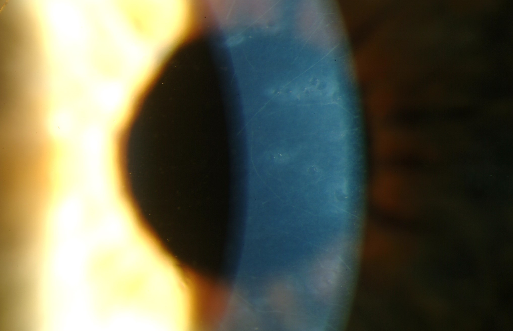
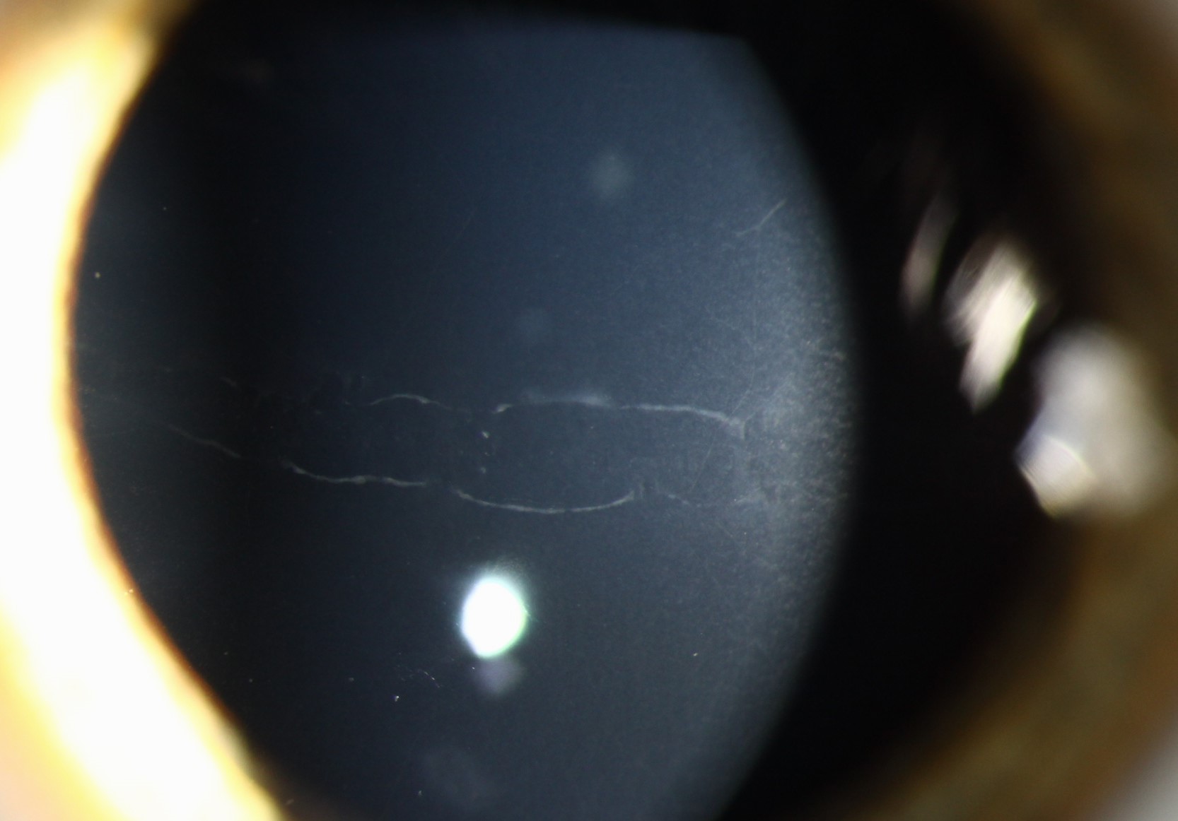
- Vesicular lesions — small, round, blister-like changes at the level of Descemet’s membrane and endothelium, often grouped in clusters
- Band-like or geographic lesions with scalloped edges, another characteristic pattern seen at the same corneal level
- Diffuse, gray, hazy opacification of Descemet’s membrane in some patients
- Most patients are asymptomatic, with normal corneal thickness and clarity to the visual axis despite the presence of these endothelial changes on careful exam
- A subset of patients develop corneal edema, sometimes significant, related to genuine endothelial dysfunction rather than simply the presence of the characteristic lesions themselves
- Iridocorneal adhesions (peripheral anterior synechiae) in more severe cases, related to the abnormal endothelial tissue extending across the angle — a feature that begins to overlap conceptually and clinically with iridocorneal endothelial (ICE) syndrome, discussed in its own dedicated article on this site, though PPCD is bilateral and inherited while ICE syndrome is characteristically unilateral and sporadic
- Secondary glaucoma, in a meaningful subset of more severely affected patients, from the same angle involvement described above
All-fit Slit-Lamp Adapter
Record and share exactly what you see at the slit lamp. One adapter fits any slit lamp or surgical microscope — and any smartphone.
From Choroida — the team behind this siteDifferential Diagnosis
- Fuchs endothelial corneal dystrophy — guttae with a “beaten metal” appearance and progressive, age-related endothelial cell loss, a different pattern and typically later onset than the vesicular/band lesions of PPCD present from a much younger age, discussed in its own dedicated article on this site
- Iridocorneal endothelial (ICE) syndrome — shares some overlapping endothelial and angle features with more severe PPCD, but is unilateral, sporadic (not inherited), and typically presents in adulthood rather than being present, even if subclinically, from a much earlier age
- Congenital hereditary endothelial dystrophy — presents with dense, diffuse corneal clouding from birth, a far more severe and immediately apparent presentation than the typically subtle, often asymptomatic findings of PPCD, discussed in its own dedicated article on this site
- Corneal guttae as a purely incidental, non-progressive finding — distinguished from true PPCD by the specific vesicular or band pattern and, when genetic testing is pursued, the relevant gene involvement
Genetics
PPCD is genetically heterogeneous, with mutations identified in several genes (including ZEB1, COL8A2, and others depending on the specific subtype), inherited in an autosomal dominant pattern with variable expressivity even within the same family.
Some relatives carrying the same mutation may show minimal, incidental findings while others develop clinically significant disease, similar in spirit to the variable expressivity discussed in this site’s coverage of dominant optic atrophy, another autosomal dominant condition where family members can be affected to different degrees.
Diagnostic Evaluation
The characteristic vesicular and band-like lesions are usually identifiable on careful slit-lamp examination with specular reflection.
Specular microscopy, where available, can further characterize the abnormal endothelial cell morphology.
Gonioscopy and intraocular pressure monitoring are important in patients with more extensive disease, given the recognized risk of secondary angle involvement and glaucoma in this subgroup, and should be incorporated into routine follow-up for these patients rather than reserved only for those with obvious symptoms.
Family screening can be informative given the autosomal dominant inheritance and variable expressivity described above.
Management
Most patients, given how often the disease remains mild and asymptomatic, require no treatment beyond periodic monitoring, including specific attention to intraocular pressure given the glaucoma risk in more severely affected individuals.
Patients who develop significant corneal edema are managed with the same general approach used for endothelial dysfunction from other causes: hypertonic saline for symptomatic relief in milder cases, and endothelial keratoplasty for more advanced, visually significant edema, following similar principles to those discussed in this site’s coverage of Fuchs endothelial dystrophy.
Secondary glaucoma related to angle involvement is managed with standard glaucoma therapy, medical or surgical depending on severity.
Family members of an affected individual may benefit from screening slit-lamp examination given the dominant inheritance pattern, even in the absence of any visual symptoms, since identifying mildly affected relatives can support informed family planning and clarify recurrence risk for future generations.


Document what you see
Two smartphone imaging tools built for everyday clinic use — one for the slit lamp, one for the fundus.
From Choroida — the team behind this siteReferences
- Weiss JS, Møller HU, Aldave AJ, et al. IC3D classification of corneal dystrophies — edition 2. Cornea.
- Krachmer JH. Posterior polymorphous corneal dystrophy: a disease characterized by epithelial-like endothelial cells which influence management and prognosis. Transactions of the American Ophthalmological Society.
- American Academy of Ophthalmology. Basic and Clinical Science Course, Section 8: External Disease and Cornea.
