Homonymous hemianopia is loss of the same half of the visual field in both eyes — both right halves or both left halves — and it localizes to the visual pathway behind the optic chiasm, in sharp contrast to the bitemporal field loss of chiasmal compression discussed in this site’s separate coverage of that condition.
Because the retrochiasmal visual pathway runs a long course from the optic tract through the thalamus and optic radiations to the occipital cortex, the exact character of a homonymous field defect — not just its presence — carries genuine localizing information about where along that pathway the responsible lesion sits.
Why “Homonymous” and Why It Localizes So Well
Beyond the chiasm, fibers carrying information from the same side of the visual field in both eyes travel together, so any lesion affecting this combined pathway on one side of the brain produces field loss on the same side (the same “half”) in both eyes simultaneously.
This is the entire basis for why a single retrochiasmal lesion produces field loss that is homonymous (matching) rather than the bitemporal or other non-matching patterns typical of lesions at or in front of the chiasm.
Congruity: The Key Localizing Concept
Congruity describes how closely the shape, size, and density of the field defect match between the two eyes.
Highly congruous defects (nearly identical in both eyes) generally localize more posteriorly, toward the occipital cortex, while less congruous (more different-appearing) defects generally localize more anteriorly, toward the optic tract or proximal optic radiations.
This general anatomic principle, taught widely in neuro-ophthalmology, is a useful rule of thumb rather than an absolute rule, but it meaningfully narrows the likely lesion location before imaging is even reviewed, giving the exam itself real predictive value.
Fundus Explorer Pro
Photograph the retinal findings described here with the phone already in your pocket — 22 D optics and built-in illumination in one handheld unit.
From Choroida — the team behind this siteLocalizing Patterns Along the Pathway
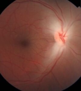
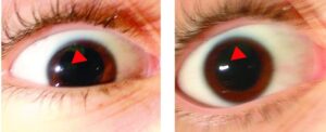
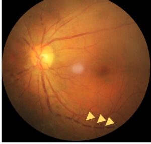
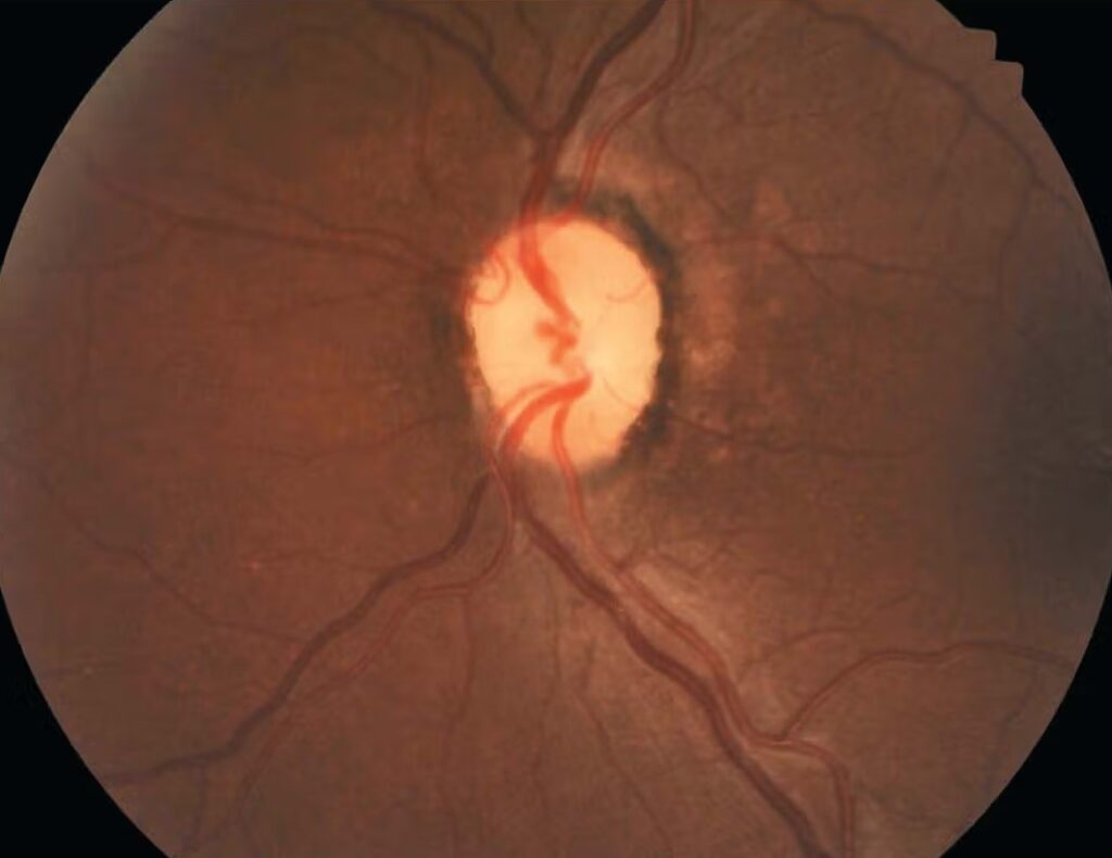
- Optic tract — incongruous homonymous hemianopia, sometimes with a relative afferent pupillary defect in the eye with the temporal field loss (since the tract carries a disproportionate share of crossed, nasal-retina fibers) and, in some cases, optic atrophy with a distinctive “bow-tie” pattern in the eye contralateral to the lesion
- Optic radiations (temporal lobe, Meyer’s loop) — a superior homonymous quadrantanopia (“pie in the sky” defect), from involvement of the inferior fibers looping through the temporal lobe
- Optic radiations (parietal lobe) — an inferior homonymous quadrantanopia, from involvement of the superior fibers taking a more direct parietal course
- Occipital cortex — highly congruous homonymous hemianopia, often with macular sparing (preservation of a small area of central field despite a dense hemianopia elsewhere), attributed to the occipital pole’s dual blood supply from both the posterior and middle cerebral arteries, which can preserve this specific cortical region even when a posterior cerebral artery stroke has damaged the rest of the visual cortex
Causes
- Stroke — the most common cause overall, particularly posterior cerebral artery territory infarction for occipital lesions
- Tumor, at any point along the retrochiasmal pathway
- Traumatic brain injury
- Demyelinating disease
- Migraine, producing transient rather than permanent homonymous field disturbance, discussed further in this site’s dedicated coverage of migraine visual aura
Clinical Presentation
Patients may report bumping into objects or people on one side, missing part of a page while reading, or difficulty with tasks requiring full peripheral awareness (driving being an important, safety-relevant example), though — similar to the pattern often seen in chiasmal compression — some patients, especially with gradual-onset lesions, are surprisingly unaware of their field loss until it is specifically tested or until a related safety incident brings it to attention.
Associated neurologic symptoms depend heavily on the underlying cause and lesion location: weakness or sensory change with a parietal lesion, memory or behavioral change with a temporal lesion, or other focal deficits corresponding to the structures near the responsible lesion, all of which contribute to localizing the lesion alongside the visual field pattern itself.
Diagnostic Evaluation
Formal visual field testing characterizes the exact pattern and congruity of the defect, providing the localizing information described above, and is generally the first, most accessible test obtained once a homonymous defect is suspected clinically.
MRI of the brain identifies the underlying structural lesion, with the region of interest guided by the specific field defect pattern found on testing.
Given how often stroke is the underlying cause, especially in an older patient with vascular risk factors and acute onset, standard stroke workup (vascular imaging, cardiac evaluation) is frequently pursued in parallel with the neuro-ophthalmic evaluation, rather than waiting for formal visual field results before initiating the broader stroke evaluation.
Management
Treatment addresses the underlying cause where treatable: acute stroke management and secondary prevention, tumor-directed therapy, or management of the underlying demyelinating process.
The visual field defect itself often does not recover once an ischemic or other structural lesion has caused permanent tissue damage, and management then shifts to visual rehabilitation strategies: compensatory scanning techniques, prism therapy in select cases, and safety counseling, particularly regarding driving, which is often restricted or requires formal evaluation once a significant, persistent homonymous field defect is confirmed.
Referral to low vision rehabilitation services, where available, can meaningfully help patients adapt daily activities and scanning strategies to their new visual field, and is worth offering proactively rather than only in response to a patient’s own request.


Document what you see
Two smartphone imaging tools built for everyday clinic use — one for the slit lamp, one for the fundus.
From Choroida — the team behind this siteReferences
- Zhang X, Kedar S, Lynn MJ, et al. Homonymous hemianopia in stroke. Journal of Neuro-Ophthalmology.
- Horton JC, Hoyt WF. The representation of the visual field in human striate cortex. Archives of Ophthalmology.
- American Academy of Ophthalmology. Basic and Clinical Science Course, Section 5: Neuro-Ophthalmology.
Test yourself
A few questions straight from this article.
-
A homonymous hemianopia localizes the lesion to which part of the visual pathway?
Loss of the same half of field in both eyes localizes to the retrochiasmal pathway, in contrast to the bitemporal field loss produced by chiasmal compression. -
In assessing a homonymous hemianopia, what does congruity describe?
Congruity is the degree to which the shape, size and density of the field defect match in the two eyes, and that match carries the localizing information. -
A highly congruous homonymous hemianopia generally localizes to which region?
Nearly identical defects in the two eyes point more posteriorly towards the occipital cortex, while less congruous defects point more anteriorly towards the optic tract or proximal radiations. -
An incongruous homonymous hemianopia with a relative afferent pupillary defect suggests a lesion in which structure?
The optic tract carries a disproportionate share of crossed nasal-retinal fibres, so tract lesions give an incongruous defect, sometimes an afferent pupillary defect in the eye with temporal field loss, and bow-tie atrophy contralaterally. -
A superior homonymous quadrantanopia, the "pie in the sky" defect, points to which lesion site?
The inferior fibres of the optic radiations loop forward through the temporal lobe as Meyer's loop, so a temporal lesion knocks out the superior field quadrant on the opposite side. -
An inferior homonymous quadrantanopia points to involvement of fibres taking which course?
The superior fibres of the optic radiations run a more direct course through the parietal lobe, so a parietal lesion produces an inferior homonymous quadrantanopia. -
Macular sparing in an occipital homonymous hemianopia is attributed to what?
The occipital pole receives blood from both the posterior and middle cerebral arteries, so a posterior cerebral artery stroke can spare that small central region while destroying the rest of the visual cortex. -
What is the most common overall cause of homonymous hemianopia?
Stroke is the most common cause overall, particularly posterior cerebral artery territory infarction when the lesion lies in the occipital cortex. -
Which investigation is generally obtained first once a homonymous field defect is suspected?
Formal perimetry is the first and most accessible test; it characterizes the exact pattern and congruity, which then guides the region of interest on brain MRI. -
Once a homonymous field defect is permanent, management shifts mainly to what?
When tissue damage is permanent, management moves to compensatory scanning techniques, prism therapy in selected cases, low vision rehabilitation referral, and counselling about driving.

Writes and reviews the clinical articles on Educate. Every post is checked against peer-reviewed sources before it goes live.