
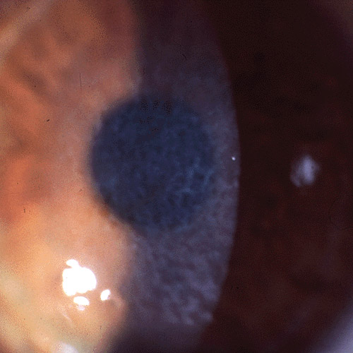
Reis-Bücklers corneal dystrophy is a rare, autosomal dominant anterior corneal dystrophy affecting Bowman’s layer, causing recurrent painful erosions and progressive superficial corneal scarring from early childhood.
It sits in a group of dystrophies once classified together as “Bowman’s layer dystrophies,” but genetic and histologic study has clarified that Reis-Bücklers and the related Thiel-Behnke dystrophy are distinct entities despite their overlapping clinical appearance.
Distinguishing the two matters mainly for prognosis and genetic counseling, since Reis-Bücklers tends to run a more severe course with earlier, more visually significant scarring.
Both conditions are rare, and most general ophthalmologists will encounter only a handful of cases across an entire career, which makes accurate classification at the time of diagnosis especially valuable for the patient’s long-term counseling.
Genetics and Pathology
Reis-Bücklers dystrophy is caused by mutations in the TGFBI gene, the same gene responsible for several other corneal stromal dystrophies including granular and lattice dystrophy, though the specific mutation and the layer affected differ.
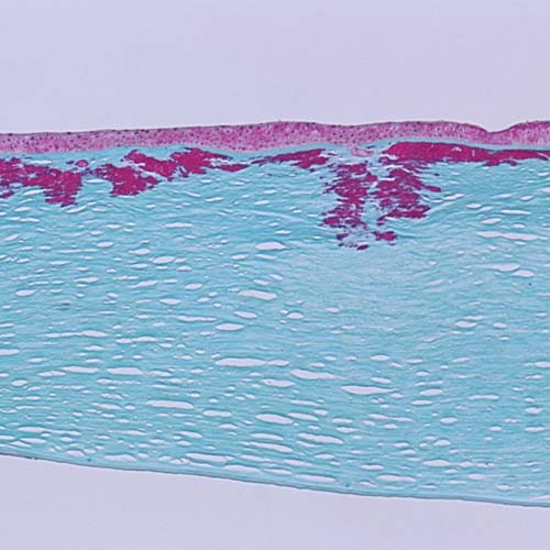
The abnormal protein deposits replace and disrupt Bowman’s layer with irregular, rod-shaped material, rather than affecting the stroma more diffusely the way granular or lattice dystrophy does.
Thiel-Behnke dystrophy, previously considered a milder variant of the same condition, is now recognized as a genetically and histologically distinct entity with a characteristic “curly fiber” pattern on electron microscopy, generally following a gentler clinical course.
Both conditions show essentially complete penetrance within affected families, so a positive family history in a child with recurrent erosions is a strong diagnostic clue even before any genetic confirmation is obtained.
Clinical Presentation
- Recurrent, painful corneal erosions beginning in early childhood, often the presenting complaint that brings the family to an ophthalmologist
- Progressive, irregular, gray-white superficial opacities concentrated in the central and paracentral cornea, coalescing over time into a honeycomb or reticular pattern
- Gradually declining vision as the opacities thicken and become more confluent, typically becoming visually significant by early-to-mid adulthood
- Reduced corneal sensation over the affected area in some patients, a secondary consequence of the recurrent epithelial disruption rather than a primary neurotrophic process
All-fit Slit-Lamp Adapter
Record and share exactly what you see at the slit lamp. One adapter fits any slit lamp or surgical microscope — and any smartphone.
From Choroida — the team behind this siteDifferential Diagnosis
Thiel-Behnke dystrophy is the closest clinical mimic, distinguished definitively by genetic testing or, when unavailable, by the generally more indolent course and characteristic ultrastructural appearance on electron microscopy.
Granular and lattice corneal dystrophy, discussed in their own dedicated articles on this site, affect the stroma rather than Bowman’s layer specifically, producing a deeper, differently distributed pattern of opacity.
Recurrent corneal erosion syndrome from other causes, including epithelial basement membrane dystrophy, needs to be considered in any patient with recurrent erosions, though the progressive Bowman’s layer opacification of Reis-Bücklers is not a feature of those other conditions.
Diagnostic Evaluation
The diagnosis is often suspected from the characteristic clinical appearance and family history, given the autosomal dominant inheritance pattern with high penetrance.
Genetic testing for the relevant TGFBI mutation can confirm the diagnosis and distinguish it definitively from Thiel-Behnke dystrophy when the clinical picture is ambiguous.
Confocal microscopy or, when tissue is available from a prior procedure, histopathology and electron microscopy can characterize the specific deposit pattern when genetic testing is not accessible.
Corneal topography is also worth obtaining as baseline documentation, since progressive Bowman’s layer scarring can eventually induce irregular astigmatism on top of the direct visual impact of the opacity itself.
Management
Recurrent erosions are managed with the same general measures used for any erosive corneal condition: lubrication, bandage contact lenses during acute episodes, and, for frequent recurrences, anterior stromal micropuncture or phototherapeutic keratectomy to promote more durable epithelial adhesion.
Phototherapeutic keratectomy, ablating the superficial opacified tissue with an excimer laser, can improve both symptoms and vision, though the dystrophy characteristically recurs in the treated area over subsequent years since the underlying genetic defect is not corrected.
Penetrating or, more often now, lamellar keratoplasty is reserved for eyes with dense, vision-limiting scarring that has progressed beyond what surface treatment can adequately address, with the same expectation that recurrence can eventually affect the graft as well.
Families benefit from understanding early that this is a lifelong, recurring condition rather than one treated definitively at a single point, since planning around that expectation shapes how aggressively surface treatment is pursued at each stage.


Document what you see
Two smartphone imaging tools built for everyday clinic use — one for the slit lamp, one for the fundus.
From Choroida — the team behind this siteReferences
- Weiss JS, Møller HU, Aldave AJ, et al. IC3D classification of corneal dystrophies — edition 2. Cornea.
- Munier FL, Korvatska E, Djemai A, et al. Kerato-epithelin mutations in four 5q31-linked corneal dystrophies. Nature Genetics.
- American Academy of Ophthalmology. Basic and Clinical Science Course, Section 8: External Disease and Cornea.
Test yourself
A few questions straight from this article.
-
Which corneal layer does Reis-Bucklers corneal dystrophy primarily affect?
Reis-Bucklers is an autosomal dominant anterior dystrophy in which abnormal deposits replace and disrupt Bowman's layer, causing recurrent erosions and superficial scarring from early childhood. -
Mutations in which gene cause Reis-Bucklers corneal dystrophy?
Reis-Bucklers results from TGFBI mutations, the same gene behind granular and lattice dystrophy, though the specific mutation and the layer affected differ. -
What does the abnormal deposit in Reis-Bucklers corneal dystrophy look like histologically?
Irregular rod-shaped protein material replaces and disrupts Bowman's layer, rather than affecting the stroma diffusely as granular or lattice dystrophy does. -
Which ultrastructural finding characterises Thiel-Behnke dystrophy on electron microscopy?
Thiel-Behnke, once thought a mild variant of Reis-Bucklers, is now a distinct entity with a characteristic curly fiber pattern on electron microscopy. -
Why does separating Reis-Bucklers from Thiel-Behnke dystrophy matter clinically?
The distinction matters mainly for prognosis and genetic counselling, since Reis-Bucklers tends to scar earlier and more significantly than Thiel-Behnke. -
How useful is a positive family history in a child with recurrent erosions suspected of Reis-Bucklers dystrophy?
Penetrance within affected families is essentially complete, so a positive family history is a strong diagnostic pointer even before genetic confirmation. -
What pattern do the corneal opacities of Reis-Bucklers dystrophy form as they coalesce?
Gray-white superficial opacities concentrated centrally and paracentrally coalesce over time into a honeycomb or reticular pattern, becoming visually significant by early-to-mid adulthood. -
Why is corneal sensation reduced in some patients with Reis-Bucklers dystrophy?
Reduced sensation over the affected area is a secondary consequence of repeated epithelial disruption, not a primary neurotrophic process. -
What is the expected course after phototherapeutic keratectomy for Reis-Bucklers dystrophy?
Excimer ablation of the opacified superficial tissue improves symptoms and vision, but the dystrophy characteristically recurs because the underlying genetic defect is untouched. -
When is keratoplasty considered in Reis-Bucklers corneal dystrophy?
Penetrating or, more often, lamellar keratoplasty is reserved for dense vision-limiting scarring, with the expectation that recurrence can eventually affect the graft too.

Writes and reviews the clinical articles on Educate. Every post is checked against peer-reviewed sources before it goes live.


