
Case Study
A 72-year-old pseudophakic man presented 10 days after trabeculectomy with sudden, painless, marked visual decline in the operated eye, reporting a “dark curtain” and profound blur.
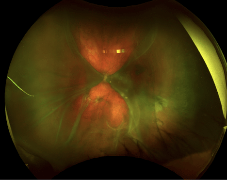
Visual acuity was light perception, intraocular pressure was 4 mmHg, and slit-lamp examination showed a deep anterior chamber with minimal inflammation but a low, diffuse filtering bleb consistent with postoperative hypotony.
Dilated fundus view was limited; B-scan ultrasonography demonstrated large bilateral convex choroidal elevations that did not insert at the optic disc and were appositional centrally—classic “kissing choroidals.
” The patient was managed with intensive topical cycloplegia and corticosteroids, cessation of aqueous suppressants, and urgent surgical drainage due to apposition and severe vision loss, with gradual anatomical improvement and partial visual recovery over follow-up.
Introduction
Choroidal detachment (also termed choroidal effusion when serous) is the accumulation of fluid or blood in the suprachoroidal space, producing smooth, dome-shaped elevations of the choroid that can extend anteriorly toward the ciliary body.
When detachments become large enough that opposing choroidal elevations meet in the mid-vitreous, the “kissing” (appositional) configuration appears, representing a severe form associated with greater risk of secondary complications and a narrower therapeutic window.
In everyday ophthalmic practice, kissing choroidals are most often encountered after glaucoma filtration or tube surgery in the setting of hypotony, but they can also occur with inflammation, trauma, infection, neoplasia, medication-related reactions, and venous congestion.
Fundus Explorer Pro
Photograph the retinal findings described here with the phone already in your pocket — 22 D optics and built-in illumination in one handheld unit.
From Choroida — the team behind this sitePathophysiology
-
A primary trigger—most commonly postoperative hypotony—lowers hydrostatic pressure within the globe and favors movement of fluid into the suprachoroidal space (a potential space between sclera and choroid).
-
As fluid accumulates, the choroid separates from the sclera and bulges inward; the effusion itself can further suppress aqueous production by affecting the ciliary body, worsening hypotony and creating a self-reinforcing cycle.
-
Detachments enlarge circumferentially and posteriorly until they become appositional (“kissing”).
-
In hemorrhagic choroidal detachment (suprachoroidal hemorrhage), rupture of choroidal/ciliary vasculature leads to rapid blood accumulation; this entity is typically more painful and carries a worse prognosis than serous effusions.
Epidemiology
Kissing choroidals are not a distinct disease but a severe configuration of choroidal detachment/effusion most frequently linked to glaucoma surgery.
Reported incidence of choroidal detachment after trabeculectomy varies widely (roughly 5%–44% in published series), reflecting heterogeneity in definitions, surgical techniques, and case mix.
Recognized risk factors in reported clinical literature include advanced age, anticoagulant use, systemic hypertension, atherosclerosis, diabetes, and trauma—particularly in the context of surgery-associated hypotony.
Clinical Features
Symptoms and severity depend on whether the detachment is serous or hemorrhagic and how extensive the apposition is.
Typical presentations include decreased vision (often profound when the macula is involved or when detachments become appositional), visual field restriction, and sometimes ocular discomfort or pain—more suggestive of hemorrhagic detachment in some reports.
Early disease may present with mild blur or peripheral shadowing, whereas appositional (“kissing”) choroidals can produce rapid, severe visual loss and may be associated with other posterior segment pathology over time.
Examination Findings
On slit-lamp examination in postoperative cases, key contextual clues include a low intraocular pressure and signs consistent with overfiltration/leak or postoperative hypotony; anterior chamber depth may be variable (from deep to shallow depending on severity and mechanism).
Fundus examination, when view is possible, may show smooth, dome-shaped, often brown elevations that are anatomically constrained by vortex vein-related adhesions, helping distinguish them from rhegmatogenous retinal detachment.
B-scan ultrasonography is especially valuable when media opacity or poor dilation limits fundoscopy: choroidal detachments appear as two convex membranes that can extend anteriorly toward the ciliary body and do not attach to the optic nerve head; appositional detachment produces the “kissing choroidal sign.”
On ultrasound, internal echogenicity may help suggest serous (more echo-lucent) versus hemorrhagic (more echo-dense) suprachoroidal contents, supporting triage and urgency.

Differential Diagnosis
-
Rhegmatogenous retinal detachment: typically forms a “V” configuration because the retina is anchored at the optic nerve head, unlike choroidal detachments, which do not insert at the disc on ultrasound.
-
Posterior scleritis: can produce exudative retinal/choroidal changes and pain; ultrasonography and clinical context help, but “kissing” apposition is more characteristic of large choroidal detachments.
-
Suprachoroidal hemorrhage vs serous choroidal effusion: both can appear as choroidal detachment; clinical pain profile and ultrasound echogenicity can help differentiate, with hemorrhagic forms generally more ominous.
-
Intraocular mass (e.g., choroidal melanoma): may mimic elevations, but detachments tend to be smooth, bilateral/symmetric in severe cases, and related to hypotony/surgery, while tumors show a localized mass effect and distinct ultrasound characteristics.
Diagnosis
Diagnosis is primarily clinical, supported by imaging—most importantly, B-scan ultrasonography when posterior segment visualization is limited.
The diagnosis of “kissing choroidals” is made when opposing choroidal detachments become appositional, typically seen on B-scan as convex choroidal elevations meeting centrally and not extending to the optic nerve head.
Determining serous versus hemorrhagic etiology relies on clinical context (hypotony after surgery vs sudden severe pain, anticoagulation, etc.) and ultrasound appearance of suprachoroidal contents.
Management
Initial management depends on cause, severity, intraocular pressure status, and whether the effusions are appositional.
Common medical steps described in postoperative hypotony-associated choroidal detachment include stopping aqueous suppressants, using cycloplegics (e.g., atropine) and topical corticosteroids, and addressing the hypotony driver (e.g., wound leak/overfiltration) while monitoring closely.
Surgical drainage is generally indicated when there is a flat/shallow anterior chamber, persistent/long-lasting effusions, significant vision loss, or appositional (“kissing”) choroidals—often treated urgently to reduce the risk of adhesions and secondary complications.
Because hemorrhagic detachments can be more vision-threatening, prompt escalation and individualized surgical timing are commonly emphasized in clinical discussions.

Prognosis
Visual prognosis varies widely and is driven by etiology (serous vs hemorrhagic), duration of apposition, degree of hypotony, and the development of secondary posterior segment complications.
Serous postoperative choroidal detachments are often reversible with appropriate management, but persistent large/appositional detachments increase the risk of structural damage and poorer visual recovery.
Hemorrhagic choroidal detachment generally carries a worse prognosis than serous effusion in published clinical descriptions.


Document what you see
Two smartphone imaging tools built for everyday clinic use — one for the slit lamp, one for the fundus.
From Choroida — the team behind this siteReferences
-
Stibbe JD, Khan NM, Doniparthi A. Kissing choroidal sign: A case report. Radiol Case Rep. 2024.
-
University of Iowa, EyeRounds.org. Atlas Entry: Choroidal Detachment: Serous, Appositional-Kissing.
-
EyeWiki. Managing Choroidal Effusions after Glaucoma Filtration Surgery. Updated 2024.
-
Glaucoma Today. Why Do Choroidals Form, and How Do You Treat Them? 2025.
-
Medscape. Choroidal Detachment: Background, Etiology, Pathophysiology.
Test yourself
A few questions straight from this article.
-
What defines the "kissing" configuration of a choroidal detachment?
When detachments grow large enough that the opposing choroidal elevations come into apposition in the mid-vitreous, the kissing configuration appears — a severe form with a narrower therapeutic window. -
In which space does fluid or blood collect in a choroidal detachment?
The suprachoroidal space is a potential space between sclera and choroid; fluid or blood entering it separates the choroid from the sclera and makes it bulge inward. -
How does a choroidal effusion perpetuate the hypotony that produced it?
The effusion affects the ciliary body and reduces aqueous production, deepening the hypotony that drew fluid into the suprachoroidal space in the first place — a self-reinforcing cycle. -
In which clinical setting are kissing choroidals most often encountered?
Kissing choroidals are most frequently seen after glaucoma filtration or tube surgery in the setting of hypotony, though inflammation, trauma, infection and neoplasia can also cause them. -
What is the reported incidence of choroidal detachment after trabeculectomy?
Published series report roughly 5% to 44%, a wide range that reflects differing definitions, surgical techniques and case mix rather than a single true figure. -
Which B-scan feature separates choroidal detachment from rhegmatogenous retinal detachment?
Choroidal detachments appear as convex membranes that extend anteriorly toward the ciliary body and do not attach at the disc, whereas the retina is anchored there and forms a V shape. -
On B-scan, what suggests haemorrhagic rather than serous suprachoroidal contents?
Serous contents are relatively echo-lucent while haemorrhagic contents are more echo-dense, and this difference helps with triage and judging urgency. -
Which clinical feature points toward a haemorrhagic rather than serous choroidal detachment?
Haemorrhagic detachment follows rupture of choroidal or ciliary vasculature, is typically more painful than a serous effusion, and carries a worse prognosis. -
Which medical step belongs in the management of hypotony-related choroidal detachment?
Medical management stops aqueous suppressants, adds cycloplegics such as atropine and topical corticosteroids, and addresses the hypotony driver such as a wound leak or overfiltration. -
Which situation makes urgent surgical drainage the right next step in choroidal detachment?
Drainage is indicated for a flat or shallow anterior chamber, persistent effusions, significant vision loss, or appositional detachments, and is done urgently to limit adhesions and secondary damage.
