
A Mittendorf dot is a small, round, white to gray opacity attached to the back surface of the lens, usually just nasal to the center and below.
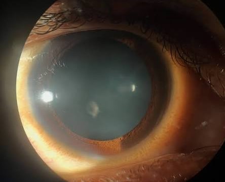
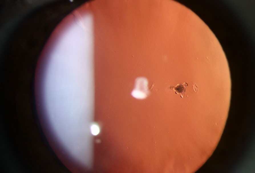
It is the place where the fetal hyaloid artery was attached to the posterior capsule, and it is a harmless developmental remnant that is found in a small proportion of otherwise normal eyes.
Where it comes from
In the fetus, the hyaloid artery runs from the optic disc through the vitreous to the back of the lens, where it supplies the tunica vasculosa lentis.
This vessel regresses before birth, and its anterior end usually leaves no trace.
Sometimes, a tiny nubbin of tissue stays on the capsule, creating the dot.
A thin strand or filament may extend from it into the vitreous.
The dot is a minimal form of the persistent fetal vasculature spectrum, which at the other end includes a retrolental plaque, a stretched ciliary process and microphthalmia (see persistent fetal vasculature).
The counterpart at the optic disc is the Bergmeister papilla (see Bergmeister papilla).
What it looks like
- A discrete, often pearl-white or grayish dot, about 0.5 to 1 mm across, on the posterior capsule
- Located slightly inferonasal to the posterior pole of the lens
- Sometimes with a fine, wispy filament that waves with eye movement
- Seen best by slit lamp with the pupil dilated and with retroillumination
It is present from birth and does not change over time.
It is found in roughly 1 to 3 percent of people, often as an incidental finding.
All-fit Slit-Lamp Adapter
Record and share exactly what you see at the slit lamp. One adapter fits any slit lamp or surgical microscope — and any smartphone.
From Choroida — the team behind this siteHow to distinguish it from other posterior lens opacities
A posterior polar cataract is a central, discoid opacity with a rosette or concentric ring appearance, and is often bilateral and familial (see posterior polar cataract).
A Mittendorf dot is small, eccentric, and does not form rings.
A posterior subcapsular cataract shows granular and vacuolated changes under the posterior capsule, usually bilateral and progressive.
The key points are that the dot is stable, small, off-axis, and unrelated to vision.
When it matters
- It does not need treatment
- It rarely affects vision, except in very rare cases when it sits in the visual axis
- It matters for cataract surgery, since the posterior capsule at that spot may be adherent or weak, and the surgeon should be aware of the possibility of a capsular defect
- A prominent filament or larger plaque should prompt a closer look for other signs of persistent fetal vasculature
What to tell the patient
Patients and parents are often worried when a spot is discovered on the lens.
They should be told that it is a leftover of normal development, that it is not a cataract, that it will not grow, and that no treatment is needed.
A note in the chart will prevent confusion at later visits.
A closer look at the remnant
The dot may appear as a single white spot, as a small cluster, or as a short thread attached to the capsule that moves slightly with eye movement. Its position, just below and nasal to the center, reflects the course of the fetal hyaloid artery from the disc to the posterior lens. It is easier to see after pupil dilation, and best seen in retroillumination, when it stands out against the red reflex as a small dark or white body.
Retroillumination also helps to distinguish it from a floating vitreous opacity, which moves independently of the lens, and from a posterior polar cataract, which has a central, rounded, plaque-like shape.
Surgical relevance
In cataract surgery, the posterior capsule at the site of a Mittendorf dot may be thin or have a defect, and the surgeon should be careful with hydrodissection and with polishing the capsule in that area. A note in the chart before surgery gives the operating team time to plan, and a mild vitrectomy setup may be kept ready if a rupture occurs.
Family and child considerations
In an infant with a dot, check the eye carefully for other signs of persistent fetal vasculature, such as a retrolental plaque, a stretched ciliary process, or a small eye. When these are absent, reassure the parents that the dot will not affect vision.
Photographs and records
Take a retroillumination photograph or a slit-lamp image of the dot when it is found. A record helps future examiners to know that the finding is old and stable, and it avoids repeat concern each time a new clinician examines the eye.
Prognosis
Excellent.
The dot has no clinical consequence in the great majority of people who have it.


Document what you see
Two smartphone imaging tools built for everyday clinic use — one for the slit lamp, one for the fundus.
From Choroida — the team behind this siteReferences
- Mann I. Developmental Abnormalities of the Eye. 2nd ed. Philadelphia: Lippincott; 1957.
- Goldberg MF. Persistent fetal vasculature (PFV): an integrated interpretation of signs and symptoms associated with persistent hyperplastic primary vitreous (PHPV). Am J Ophthalmol. 1997;124:587-626.
- Pollard ZF. Persistent hyperplastic primary vitreous: diagnosis, treatment and results. Trans Am Ophthalmol Soc. 1997;95:487-549.
- Duke-Elder S. System of Ophthalmology. Vol III: Normal and Abnormal Development. London: Kimpton; 1964.
