
Case Study
A 56-year-old female with a history of high myopia and recent trauma presented to an emergency department with sudden onset of floaters, flashes of light, and visual field loss in her left eye.

The patient reported that the symptoms began shortly after accidentally hitting her eye on the corner of a door. She denied any pain or previous history of ocular surgeries or retinal issues.
Examination Results:
- Visual Acuity: 20/40 in the right eye, 20/200 in the left eye.
- Fundus Examination: The left eye revealed a retinal tear along the periphery with evidence of vitreous hemorrhage. There was also a visible avulsion of the vitreous base, with the vitreous being detached from its normal attachment at the ora serrata.
- B-scan Ultrasonography: Confirmed vitreous base avulsion with vitreous hemorrhage and partial retinal detachment.
- Intraocular Pressure (IOP): 14 mmHg in both eyes.
A diagnosis of Vitreous Base Avulsion secondary to blunt ocular trauma was made.
Vitreous Base Avulsion Disease Entity
Vitreous base avulsion is a serious ocular condition characterized by the separation of the vitreous base from its attachment at the ora serrata.
This detachment often results from severe blunt trauma to the eye, leading to the disruption of the structural integrity of the vitreoretinal interface.
A vitreous base avulsion is frequently associated with retinal tears, vitreous hemorrhage, and an increased risk of retinal detachment.
Pathophysiology
The vitreous base is a critical region where the vitreous gel adheres firmly to the retina and the pars plana of the ciliary body, extending about 2 mm anterior to the ora serrata and 4 mm posterior to it.
During blunt ocular trauma, the forces exerted on the eye can cause significant shearing stress at the vitreous base, leading to its avulsion.
This can result in the vitreous pulling away from the retina, creating retinal tears or holes and leading to vitreous hemorrhage.
When the vitreous base is avulsed, the mechanical disruption at the site can lead to retinal tears, particularly at the peripheral retina, which is already susceptible due to its thinner structure.
The subsequent vitreous hemorrhage may obscure the view of the retina, making diagnosis and management more challenging.
The presence of vitreous base avulsion significantly increases the risk of retinal detachment, which can lead to severe visual impairment if not promptly treated.
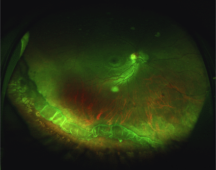
Vitreous base avulsion Epidemiology
Vitreous base avulsion is relatively rare but is most commonly seen in individuals who have sustained significant blunt trauma to the eye, such as from motor vehicle accidents, sports injuries, or physical assaults.
It is more frequently observed in patients with predisposing factors such as high myopia, previous eye surgeries, or pre-existing retinal degenerations.
The condition can occur at any age but is more common in younger, active individuals who are more likely to sustain blunt trauma.
The incidence of vitreous base avulsion may be underreported due to the subtlety of its presentation and the challenges in diagnosing it, particularly when associated with dense vitreous hemorrhage.
Vitreous base avulsion Clinical Features
Patients with vitreous base avulsion typically present with symptoms related to the traumatic disruption of the vitreoretinal interface:
- Sudden Visual Field Loss: Often described as a curtain or shadow over the vision, usually corresponding to the area of retinal detachment.
- Floaters: Due to vitreous hemorrhage or posterior vitreous detachment, patients may notice an increase in floaters.
- Photopsia: Flashes of light are common, resulting from tractional forces on the retina.
- Blunt Trauma History: A history of significant ocular trauma is almost always present.
Examination Findings
- Fundus Examination: Key findings include peripheral retinal tears, vitreous hemorrhage, and detachment of the vitreous base from the ora serrata. The presence of blood in the vitreous cavity may complicate the visualization of the retina.
- B-scan Ultrasonography: Essential in cases where vitreous hemorrhage obscures the retina, B-scan ultrasound can confirm the diagnosis by visualizing the vitreous base avulsion, retinal tears, and the extent of retinal detachment.
- Optical Coherence Tomography (OCT): May reveal subtle retinal changes and provide detailed imaging of the vitreoretinal interface.

Differential Diagnosis
The differential diagnosis of vitreous base avulsion includes other traumatic and non-traumatic retinal conditions:
- Posterior Vitreous Detachment (PVD): Although more common and less severe, PVD can also cause floaters and flashes but lacks the traumatic etiology and association with peripheral retinal tears.
- Retinal Detachment: A primary retinal detachment without vitreous base avulsion may present similarly but does not involve the specific avulsion of the vitreous base.
- Vitreous Hemorrhage: Vitreous hemorrhage without an associated vitreous base avulsion may occur in conditions like diabetic retinopathy or retinal vein occlusion.
- Choroidal Rupture: Traumatic choroidal rupture may present with visual disturbances and hemorrhage but typically involves the choroid rather than the vitreous base.
Vitreous base avulsion Diagnosis
Diagnosis of vitreous base avulsion is based on clinical examination and imaging studies:
- Fundus Examination: Visualization of the avulsed vitreous base and associated retinal pathology is crucial.
- B-scan Ultrasonography: Provides critical information in cases where vitreous hemorrhage obscures the view, confirming the diagnosis and assessing the extent of damage.
- Optical Coherence Tomography (OCT): May be used for detailed imaging of the vitreoretinal interface, although its utility may be limited in acute cases with significant hemorrhage.
Vitreous base avulsion Management
Management of vitreous base avulsion focuses on preventing retinal detachment and managing associated complications:
- Vitrectomy: Surgical intervention, often involving pars plana vitrectomy, may be necessary to remove vitreous hemorrhage, repair retinal tears, and reattach the retina.
- Retinopexy: Laser retinopexy or cryotherapy may be used to secure the retina around the tear and prevent detachment.
- Scleral Buckling: In some cases, scleral buckling may be employed to support the retina and reduce traction on the vitreous base.
- Regular Monitoring: Ongoing follow-up is essential to monitor for any signs of retinal detachment or further vitreous complications.

Prognosis
The prognosis for patients with vitreous base avulsion varies depending on the severity of the initial trauma and the promptness of treatment.
If retinal detachment is promptly addressed, the visual prognosis can be favorable. However, if left untreated, vitreous base avulsion can lead to complete retinal detachment and significant, potentially permanent, vision loss.
Prevention
Preventing vitreous base avulsion involves minimizing the risk of ocular trauma:
- Protective Eyewear: Especially important for individuals involved in high-risk activities or contact sports.
- Prompt Treatment of Ocular Injuries: Immediate evaluation and management of any ocular trauma are critical to prevent complications like vitreous base avulsion.
- Education: Raising awareness about the importance of eye protection and prompt medical evaluation following trauma can reduce the incidence of this condition.
References
- Blanch, R. J., & Mohammed, Q. (2013). Traumatic vitreous base avulsion: A case series and review of the literature.
- Kuhn, F., & Pieramici, D. J. (2002). Ocular trauma: Principles and practice. New York: Thieme.
- Coleman, D. J., & Trokel, S. (1969). Direct-recorded intraocular hydrostatic pressures in human eyes: Experimental findings. Archives of Ophthalmology, 82(5), 638-644.
- Scott, I. U., Flynn Jr, H. W., & Feuer, W. J. (1997). Management of traumatic retinal detachment associated with vitreous base avulsion: Surgical outcomes and prognostic factors.
- Khalil, H., & Leonard, M. (2010). Vitreous base avulsion in sports-related eye injuries. British Journal of Sports Medicine, 44(8), 568-571.


Document what you see
Two smartphone imaging tools built for everyday clinic use — one for the slit lamp, one for the fundus.
From Choroida — the team behind this siteTest yourself
A few questions straight from this article.
-
What defines vitreous base avulsion?
Vitreous base avulsion is the separation of the vitreous base from its attachment at the ora serrata, usually after severe blunt trauma, with disruption of the vitreoretinal interface. -
To which structures does the vitreous base adhere most firmly?
The vitreous base is the region where the vitreous gel adheres firmly to the retina and to the pars plana of the ciliary body, which is why trauma there disrupts the vitreoretinal interface. -
What mechanical event produces vitreous base avulsion during blunt ocular trauma?
Blunt trauma generates significant shearing stress at the vitreous base, which avulses it and pulls the vitreous away from the retina, creating tears and vitreous haemorrhage. -
Why do retinal tears from vitreous base avulsion arise particularly in the peripheral retina?
Mechanical disruption at the avulsed vitreous base tends to tear the peripheral retina, which is already susceptible because of its thinner structure. -
The presence of a vitreous base avulsion most importantly raises the risk of which complication?
Vitreous base avulsion significantly increases the risk of retinal detachment, which causes severe visual impairment unless treated promptly. -
Which investigation is essential when vitreous haemorrhage obscures the retina after suspected vitreous base avulsion?
Where vitreous haemorrhage blocks the view, B-scan ultrasonography is essential: it confirms the vitreous base avulsion and shows retinal tears and the extent of any detachment. -
A patient with vitreous base avulsion describes a curtain across the vision. What does this correspond to?
Sudden visual field loss described as a curtain or shadow usually corresponds to the area of retinal detachment in these eyes. -
Which feature separates simple posterior vitreous detachment from vitreous base avulsion?
Posterior vitreous detachment is commoner and less severe and can also cause floaters and flashes, but it lacks the traumatic aetiology and the association with peripheral retinal tears seen in vitreous base avulsion. -
Which procedure is used to secure the retina around a tear in vitreous base avulsion?
Laser retinopexy or cryotherapy is applied around the retinal tear to secure the retina and prevent progression to detachment. -
What is the visual prognosis after vitreous base avulsion?
Outcome depends on the severity of the trauma and how quickly treatment is given: promptly treated retinal detachment can do well, while untreated avulsion can progress to complete detachment and permanent vision loss.
