
Molluscum contagiosum is a common poxvirus infection of the skin that, when it occurs on or near the eyelid margin, can produce a chronic, often stubbornly persistent follicular conjunctivitis that is frequently misdiagnosed and mistreated as allergic or viral conjunctivitis until someone specifically looks for the small, characteristic skin lesion actually driving it.
Recognizing the lesion itself — rather than just treating the conjunctival inflammation it causes — is the key to resolving the problem, and once it is correctly identified, the underlying diagnosis and treatment approach usually become straightforward.
Pathogenesis
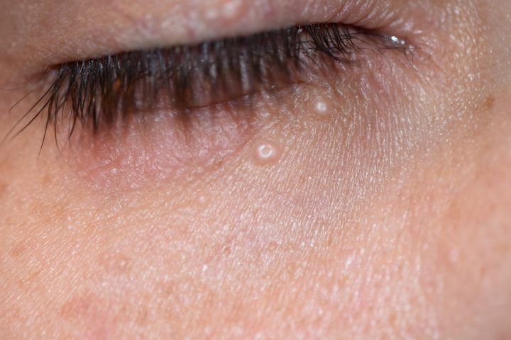
The molluscum contagiosum virus infects epidermal keratinocytes, producing small, discrete papules with a central core of infectious viral particles.
When a lesion sits on or near the eyelid margin, this viral material sheds continuously into the tear film with every blink, producing a chronic toxic and follicular immune reaction on the conjunctiva that persists for as long as the lesion remains untreated.
The conjunctivitis is a downstream consequence of ongoing viral shedding, not a separate process that needs its own independent treatment.
This distinction — conjunctivitis as a secondary effect rather than the primary disease — is the single most important conceptual point for correctly managing this condition, since treatments aimed only at the eye surface will continue to fail for as long as the actual lesion remains untreated.
Populations at Risk
- Children — the most commonly affected group in the general population, often with lesions elsewhere on the body from typical skin-to-skin spread
- Immunocompromised patients, particularly those with HIV/AIDS, where lesions can be larger, more numerous, and more treatment-resistant than in immunocompetent patients
- Sexually active young adults, when lesions occur in the periocular region as part of broader genital/perigenital involvement
In an adult presenting with new, multiple, or unusually large molluscum lesions, with periocular involvement, an underlying immunocompromising condition should be considered and, where appropriate, tested for, because the presentation can differ meaningfully from the typical, self-limited childhood pattern, both in extent and in how readily it responds to standard treatment.
All-fit Slit-Lamp Adapter
Record and share exactly what you see at the slit lamp. One adapter fits any slit lamp or surgical microscope — and any smartphone.
From Choroida — the team behind this siteClinical Presentation
Patients (or, more often, a parent) present with chronic, unilateral, treatment-resistant red eye, mild irritation, and mucoid discharge, often having already tried topical antibiotics or antihistamines without improvement.
That lack of response to standard conjunctivitis treatment is itself a useful diagnostic clue pointing toward an unusual cause like molluscum.
The chronicity, frequently lasting weeks to months before the diagnosis is made, distinguishes it from the more acute course of typical viral or bacterial conjunctivitis.
Because the eyelid margin is not always the first area a clinician inspects during a routine conjunctivitis evaluation, deliberately examining the full lid margin, including everting the lid when necessary, is an important step whenever a conjunctivitis has proven unexpectedly persistent or unresponsive to standard treatment.
Exam Findings
- One or more small (typically 1–3 mm), dome-shaped, pearly or flesh-colored papules with a characteristic central umbilication, located on or near the eyelid margin
- A follicular reaction on the ipsilateral conjunctiva, from the chronic viral shedding described above
- Mild to moderate conjunctival injection
- Absence of significant preauricular lymphadenopathy, which, when present, should prompt reconsideration of the diagnosis toward viral or chlamydial conjunctivitis instead
- Superficial punctate keratitis or, in more longstanding cases, mild corneal pannus from chronic irritation
Differential Diagnosis
- Adenoviral (epidemic) keratoconjunctivitis — usually more acute in onset, with preauricular lymphadenopathy and a shorter overall course
- Adult inclusion (chlamydial) conjunctivitis — chronic follicular conjunctivitis as well, but without a visible eyelid margin lesion, and with a relevant sexual history
- Verruca (viral wart) — a different virus (human papillomavirus) producing a papillomatous rather than dome-shaped, umbilicated lesion
- Chalazion or hordeolum — inflammatory eyelid lumps without the characteristic central umbilication or the associated chronic follicular conjunctivitis
Diagnosis
The diagnosis is made clinically based on the characteristic appearance of the lesion, particularly the central umbilication, which is specific enough that biopsy is rarely needed in a typical presentation.
Biopsy is reasonable when the lesion appearance is atypical, when it fails to respond to treatment as expected, or when there is concern for an underlying immunocompromising condition given the atypical presentation that can sometimes accompany it.
Management
Removal of the causative lesion — whether by simple excision, curettage, or cryotherapy — resolves the associated conjunctivitis, generally within a couple of weeks, once the source of ongoing viral shedding is eliminated.
This is the definitive treatment; topical medications directed at the conjunctivitis itself without addressing the lesion typically fail or provide only partial, temporary relief.
In immunocompetent children with small, few lesions and minimal symptoms, observation is a reasonable alternative, because molluscum contagiosum is generally self-limited and will often resolve on its own over months.
However, this comes at the cost of prolonged conjunctival irritation in the interim if the eyelid lesion is actively shedding virus.
In immunocompromised patients, lesions may be more numerous, larger, and less likely to resolve without treatment, and more aggressive or repeated intervention, along with attention to the underlying immune status, is often needed for durable resolution.
Coordination with the patient’s HIV or other immunosuppression management team is appropriate in these more resistant cases, since improving the underlying immune status can itself meaningfully affect how well the periocular lesions ultimately respond to local treatment.


Document what you see
Two smartphone imaging tools built for everyday clinic use — one for the slit lamp, one for the fundus.
From Choroida — the team behind this siteReferences
- Charteris DG, Bonshek RE, Tullo AB. Ophthalmic molluscum contagiosum: clinical and immunopathological features. British Journal of Ophthalmology.
- Robinson MR, Udell IJ, Garber PF, et al. Molluscum contagiosum of the eyelids in patients with acquired immune deficiency syndrome. Ophthalmology.
- American Academy of Ophthalmology. Basic and Clinical Science Course, Section 8: External Disease and Cornea.
Test yourself
A few questions straight from this article.
-
Which organism causes molluscum contagiosum of the eyelid?
The molluscum contagiosum virus is a poxvirus that infects epidermal keratinocytes, producing small discrete papules with a central core of infectious viral particles. -
How does an eyelid margin molluscum lesion produce conjunctivitis?
Viral material sheds into the tear film with every blink, producing a chronic toxic and follicular immune reaction that persists for as long as the lesion is left untreated. -
What does an eyelid molluscum contagiosum lesion look like on examination?
Lesions are typically 1-3 mm, dome-shaped and pearly or flesh-coloured, with the characteristic central umbilication that makes the diagnosis a clinical one. -
What should significant preauricular lymphadenopathy suggest in suspected eyelid molluscum contagiosum?
Molluscum-related conjunctivitis characteristically lacks significant preauricular lymphadenopathy, so finding it should push the diagnosis toward a viral or chlamydial cause instead. -
Which presenting pattern should raise suspicion for eyelid molluscum contagiosum?
Patients typically present after weeks to months of unilateral irritation and mucoid discharge, having already failed topical antibiotics or antihistamines. That lack of response is itself the clue. -
What should new, multiple or unusually large periocular molluscum lesions in an adult prompt?
Immunocompromised patients, particularly with HIV/AIDS, develop larger, more numerous and more treatment-resistant lesions, so the presentation differs meaningfully from the self-limited childhood pattern. -
Which feature distinguishes a verruca from eyelid molluscum contagiosum?
Verruca is caused by human papillomavirus and produces a papillomatous lesion, lacking the dome-shaped, centrally umbilicated appearance of molluscum contagiosum. -
When is biopsy reasonable in suspected eyelid molluscum contagiosum?
Central umbilication is specific enough that biopsy is rarely needed in a typical case; it is reserved for atypical appearance, unexpected treatment failure, or concern about immunocompromise. -
What is the definitive treatment for molluscum-associated conjunctivitis?
Excision, curettage or cryotherapy eliminates the source of viral shedding and the conjunctivitis generally resolves within a couple of weeks. Drops aimed at the eye surface alone typically fail. -
When is observation a reasonable option in eyelid molluscum contagiosum?
Molluscum is generally self-limited and often resolves over months, so observation is reasonable when symptoms are minimal, though it means prolonged conjunctival irritation while the lesion keeps shedding.
