
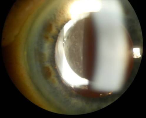
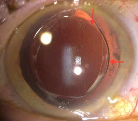
Negative dysphotopsia is a genuinely puzzling visual complaint following otherwise uncomplicated, technically successful cataract surgery: a dark, crescent-shaped shadow, typically in the temporal peripheral visual field, that can be persistently bothersome to a subset of patients despite a well-centered intraocular lens, a clear visual axis, and excellent measured visual acuity.
Why This Complication Is So Frustrating
Negative dysphotopsia occurs in eyes with no identifiable surgical error, a properly positioned intraocular lens, and no other explanation for the symptom on standard examination, which makes it a particularly difficult complication to explain to patients and, historically, a difficult one to manage given uncertainty about its precise underlying cause.
Its symptomatic profile, an annoying dark shadow rather than reduced visual acuity, also means that standard visual acuity testing, the metric most surgeons and patients focus on as the marker of surgical success, does not capture the impact of this complication at all.
Proposed Mechanisms
Several mechanisms have been proposed, and current understanding suggests the phenomenon likely results from a combination of factors related to how light interacts with the edge of the intraocular lens optic and the surrounding anatomy.
- Light rays passing near the sharp edge of a modern square-edge intraocular lens optic (a design feature intentionally used to reduce posterior capsular opacification) can be refracted in a way that creates a gap in retinal illumination, perceived as a shadow
- The specific anatomic relationship between the lens position, the iris, and the nasal retina appears to influence whether and how strongly a given eye experiences the phenomenon
- Some degree of angle kappa (the angular relationship between the visual and pupillary axes) may contribute to individual variation in symptom presence and severity
All-fit Slit-Lamp Adapter
Record and share exactly what you see at the slit lamp. One adapter fits any slit lamp or surgical microscope — and any smartphone.
From Choroida — the team behind this siteClinical Presentation
- A dark, often crescent or arc-shaped shadow in the temporal peripheral visual field, generally most noticeable in bright light and in the periphery of vision rather than centrally
- Onset is typically within the first days after surgery
- Visual acuity testing is normal, and the ocular examination, including intraocular lens position, is unremarkable, which is part of what makes this condition so puzzling for both patient and surgeon
- Symptoms improve or resolve spontaneously in a substantial proportion of patients over the weeks to months following surgery, thought to reflect a degree of neuroadaptation
Natural History
A meaningful proportion of patients experience spontaneous improvement or resolution of negative dysphotopsia symptoms over time, generally within the first several months after surgery, which supports an initial period of observation and reassurance as a reasonable first step for many patients before considering more active intervention.
Management
Observation and Reassurance
Given the tendency toward spontaneous improvement, initial management for most patients centers on reassurance and observation, along with clear counseling that this is a recognized, generally benign, though sometimes persistent, phenomenon.
Nonsurgical Measures
Some patients find partial relief from stronger reading glasses or other approaches attempting to fill in the peripheral field gap, though these measures are generally only partially effective.
Surgical Options for Persistent, Bothersome Symptoms
For patients with persistent, significantly bothersome symptoms not improving with observation, several surgical approaches have been described, aiming to alter the relevant optical geometry at the lens edge.
- Reverse optic capture, in which the intraocular lens optic is repositioned so that the anterior capsule edge sits behind the optic rather than in front of it, has shown good success in reducing symptoms in reported case series
- Piggyback intraocular lens placement, adding a second lens in the ciliary sulcus, can alter the optical pathway sufficiently to relieve symptoms in some patients
- Exchange to a different intraocular lens design, particularly one without the same sharp anterior edge profile, or repositioning the existing lens, are additional options considered in refractory cases
Prognosis
Most patients with negative dysphotopsia experience meaningful improvement over time, whether through spontaneous neuroadaptation or, for the smaller subset with persistent, significant symptoms, through targeted surgical intervention.
Preoperative counseling about this recognized, if uncommon, phenomenon, particularly for patients undergoing surgery in their better-seeing or only functional eye, helps set realistic expectations, given that a small proportion of patients will experience this frustrating symptom despite an otherwise textbook-successful cataract surgery.


Document what you see
Two smartphone imaging tools built for everyday clinic use — one for the slit lamp, one for the fundus.
From Choroida — the team behind this siteReferences
- Davison JA. Positive and negative dysphotopsia in patients with acrylic intraocular lenses. J Cataract Refract Surg. 2000;26:1346-1355.
- Masket S, Fram NR. Pseudophakic negative dysphotopsia: surgical management and new theory of etiology. J Cataract Refract Surg. 2011;37:1199-1207.
- Holladay JT, Simpson MJ. Negative dysphotopsia: causes and rationale for prevention and treatment. J Cataract Refract Surg. 2017;43:263-275.
- Cooke DL, Kasko S, Platt LO. Resolution of negative dysphotopsia after laser anterior capsulotomy. J Cataract Refract Surg. 2013;39:1107-1109.
