
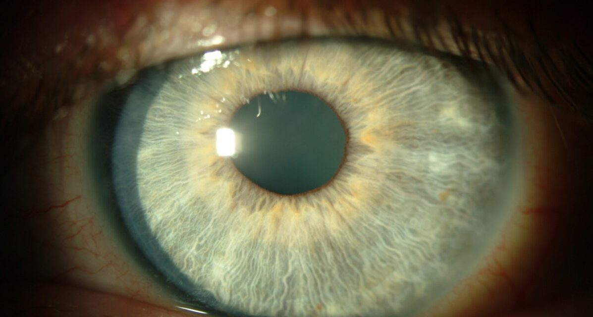
Thygeson Superficial Punctate Keratitis is the corneal disease that keeps coming back long after everyone expected it to settle.
Patients complain bitterly of grittiness and light sensitivity, yet the eye looks almost white and quiet.
The only clue is a scattering of tiny grey dots on the cornea that flare, fade, and return over months or years.
For clinicians, recognizing Thygeson Superficial Punctate Keratitis (TSPK) matters because it is neither infective nor dangerous, but it is repeatedly mistaken for viral keratitis and treated with the wrong drugs.
A calm eye with symptoms out of proportion to the signs, running a relapsing course, should always raise the possibility.
What Is Thygeson Superficial Punctate Keratitis?
TSPK is a chronic, bilateral, recurrent epithelial keratitis of unknown cause, first described by Phillips Thygeson in 1950.
Its defining features are:
- Coarse, grey-white, slightly raised intraepithelial corneal opacities
- A bilateral, relapsing-and-remitting course over years
- Symptoms that are worse than the modest clinical signs
- A quiet eye with little or no conjunctival reaction
It is a disease of the corneal epithelium alone, which is why it heals without scarring.
Epidemiology
The condition is uncommon and affects a broad age range.
- Can present at any age, but often in young to middle-aged adults
- Usually bilateral, though it may be asymmetric
- An association with HLA-DR3 has been reported
Because the course spans years, many patients accumulate several incorrect diagnoses before the pattern becomes clear.
All-fit Slit-Lamp Adapter
Record and share exactly what you see at the slit lamp. One adapter fits any slit lamp or surgical microscope — and any smartphone.
From Choroida — the team behind this sitePathogenesis
The cause remains genuinely unknown.
- A viral trigger has long been suspected but never confirmed
- An immune-mediated mechanism is supported by the response to corticosteroids and ciclosporin
- The HLA-DR3 association hints at a genetic predisposition
Whatever the trigger, the process stays confined to the epithelium and spares the stroma.
Clinical Presentation
Symptoms
The symptoms are strikingly out of proportion to the appearance.
- Foreign-body sensation and grittiness
- Photophobia and tearing
- Intermittent blurring during flares
- A relapsing course with symptom-free intervals
Examination Findings
The diagnosis is made at the slit lamp.
- Coarse punctate epithelial opacities: grey-white, slightly elevated, often central
- Lesions that stain with fluorescein during active phases and fade to faint grey dots in quiescence
- A quiet eye with minimal or absent conjunctival injection
- No stromal infiltrate and no anterior chamber reaction
- Normal corneal sensation
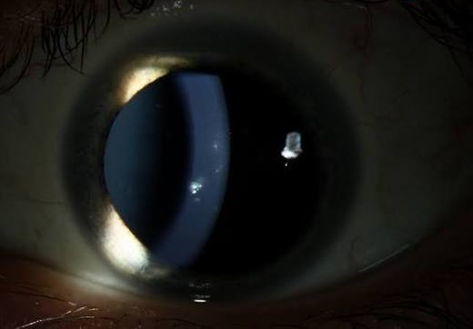
The mismatch between marked symptoms and a nearly white eye is the single most useful clue.
Why Thygeson Keratitis Is So Often Misdiagnosed
Almost every wrong turn follows the same reasoning.
- The epithelial dots are mistaken for herpes simplex keratitis, and antivirals are started needlessly
- It is confused with adenoviral keratoconjunctivitis, though there is no red eye or preauricular node
- It is labelled simple dry eye, and the relapsing pattern is overlooked
- Prolonged steroid use is started without a plan, risking dependence and complications
A bilateral, recurrent punctate keratitis in a quiet eye that never scars should point to TSPK rather than infection.
Diagnostic Evaluation
The diagnosis is clinical and rests on the pattern over time.
Clinical Assessment
- Slit-lamp examination with fluorescein to characterize the epithelial lesions
- Assessment of corneal sensation, which is preserved
- Documentation of the bilateral, relapsing course
When to Investigate Further
- Viral swabs or scrapings are reserved for atypical or unilateral cases to exclude herpes
- No specific laboratory test confirms TSPK; it remains a clinical diagnosis
Serial slit-lamp photographs are invaluable for tracking a disease defined by its behaviour over time.
Differential Diagnosis
Several epithelial keratopathies can look similar:
- Herpes simplex epithelial keratitis — true dendrites and reduced corneal sensation
- Adenoviral keratoconjunctivitis — red eye, follicles and a preauricular node
- Staphylococcal marginal keratitis — peripheral infiltrates with a clear zone
- Dry eye and exposure keratopathy — inferior punctate staining without raised dots
- Infective keratitis — an infiltrate, pain and an inflamed eye
The combination of coarse intraepithelial dots, a quiet eye, and a chronic relapsing course sets TSPK apart.
Management
Treatment is aimed at comfort during flares, not cure.
- Lubricants are first-line for mild disease
- Low-potency topical corticosteroids shorten flares but should be used in short, tapering courses to avoid dependence
- Topical ciclosporin is a useful steroid-sparing option for frequent recurrences
- A therapeutic soft contact lens can relieve symptoms during severe episodes
- Antivirals and antibiotics are ineffective and should be avoided
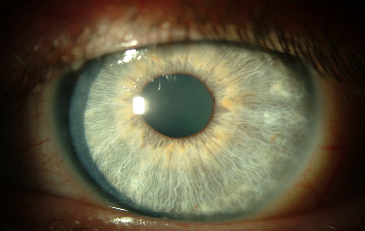
The guiding principle is to treat symptoms with the least medication needed and to resist open-ended steroid use.
Prognosis
The long-term outlook is excellent.
- Vision is preserved because the stroma is spared and scarring is rare
- The disease is self-limiting, though it may recur for years or decades
- Most patients need reassurance and intermittent treatment rather than continuous therapy
In practice, the greatest service is making the diagnosis, stopping unnecessary antivirals, and reassuring the patient about the benign course.
Would you like to document corneal findings like Thygeson dots with your smartphone?


Document what you see
Two smartphone imaging tools built for everyday clinic use — one for the slit lamp, one for the fundus.
From Choroida — the team behind this siteReferences
- Thygeson P. “Superficial Punctate Keratitis.” JAMA. 1950;144(18):1544–1549.
- Nagra PK, Rapuano CJ, Laibson PR, et al. “Thygeson’s Superficial Punctate Keratitis: Ten Years’ Experience.” Ophthalmology. 2004;111(1):34–37.
- Tabbara KF, Ostler HB, Dawson C, Oh J. “Thygeson’s Superficial Punctate Keratitis.” Ophthalmology. 1981;88(1):75–77.
- Reinhard T, Sundmacher R. “Topical Ciclosporin A in Thygeson’s Superficial Punctate Keratitis.” Graefe’s Archive for Clinical and Experimental Ophthalmology. 1999;237(2):109–112.
- American Academy of Ophthalmology. “Thygeson’s Superficial Punctate Keratitis.” EyeWiki.
Test yourself
A few questions straight from this article.
-
Thygeson superficial punctate keratitis is best described as which kind of corneal disease?
TSPK is a chronic, bilateral, relapsing epithelial keratitis of unknown aetiology, first described by Phillips Thygeson in 1950. -
Which clinical mismatch is the single most useful clue to Thygeson superficial punctate keratitis?
Patients complain bitterly of grittiness and photophobia while the eye looks quiet with little or no conjunctival reaction, a mismatch that should raise TSPK. -
Which HLA type has been reported in association with Thygeson superficial punctate keratitis?
An HLA-DR3 association has been reported and hints at a genetic predisposition, although the underlying cause of TSPK remains unknown. -
Why does Thygeson superficial punctate keratitis characteristically heal without corneal scarring?
TSPK is a disease of the corneal epithelium alone; the stroma is spared, so vision is preserved and scarring is rare. -
What do the corneal lesions of Thygeson superficial punctate keratitis look like at the slit lamp?
The dots are coarse, grey-white, slightly elevated and often central; they stain with fluorescein during flares and fade to faint grey specks in quiescence. -
Which examination finding is preserved in Thygeson superficial punctate keratitis and helps exclude herpes simplex keratitis?
Corneal sensation is normal in TSPK, whereas herpes simplex epithelial keratitis shows true dendrites with reduced corneal sensation. -
Which accompanying signs point to adenoviral keratoconjunctivitis rather than Thygeson keratitis?
Adenoviral disease produces a red eye with follicles and a preauricular node, none of which accompany the quiet eye of TSPK. -
What is the first-line treatment for mild Thygeson superficial punctate keratitis?
Lubricants are first-line for mild disease; antivirals and antibiotics are ineffective in TSPK and should be avoided. -
Which steroid-sparing topical agent is useful for frequently recurring Thygeson superficial punctate keratitis?
Topical ciclosporin is a useful steroid-sparing option for frequent recurrences, avoiding the dependence and complications of open-ended steroid use. -
What is the long-term visual prognosis in Thygeson superficial punctate keratitis?
The stroma is spared and scarring is rare, so vision is preserved; the disease is self-limiting but may relapse over years or decades.
