
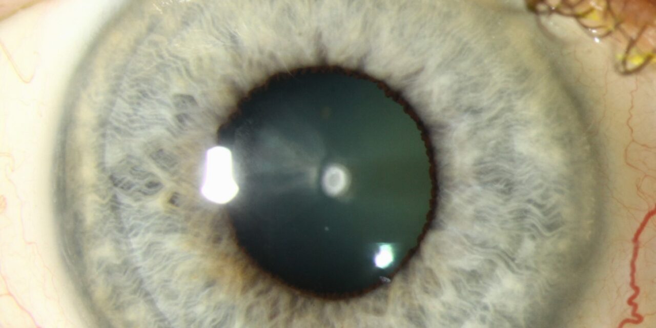
Anterior polar cataract is a small, sharply demarcated white opacity at the front of the lens, sitting just beneath the anterior capsule at the center of the pupillary axis.
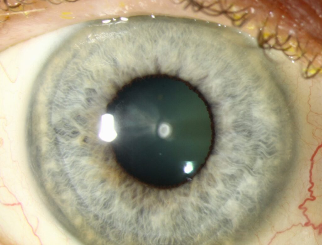
It is congenital, usually stable rather than progressive, and — in sharp contrast to its posterior counterpart — carries none of the surgical risk that makes posterior polar cataract so notable, because the capsule beneath it is normal rather than thinned or absent.
It is also one of the more commonly encountered congenital lens opacities, frequently picked up as an incidental finding during a routine pediatric red reflex screen rather than because a parent noticed any visual problem.
Pathogenesis
The lesion arises from a localized failure of normal separation between the lens vesicle and the overlying surface ectoderm during early embryonic development, leaving a small island of abnormal epithelial or fibrous tissue adherent to the central anterior capsule.
Most cases are sporadic, though autosomal dominant inheritance occurs in some families, usually with a small, stable, non-progressive lesion passed down without much variability in severity between affected relatives.
A distinct, more aggressive variant — anterior pyramidal cataract — presents as a cone-shaped opacity that can protrude through the pupil into the anterior chamber and is more often associated with progression and anterior uveitis-like inflammatory changes; this variant is managed differently and with more urgency than the simple, flat anterior polar form.
Because the two forms share a similar location and can look superficially alike on a brief exam, correctly distinguishing a flat, stable anterior polar opacity from the raised, protruding pyramidal variant at the initial visit meaningfully affects how closely the child needs to be followed afterward.
Clinical Presentation
Because the opacity is typically small (often 1 to 3 mm) and centered precisely on the visual axis, its visual impact depends heavily on size: many are visually insignificant and discovered incidentally, while larger lesions can cause measurable reduction in acuity and, in a young child, a genuine risk of amblyopia if left uncorrected.
Unlike posterior subcapsular or nuclear cataracts, anterior polar cataract does not typically progress over a patient’s lifetime, so a stable, small lesion identified in childhood usually remains essentially unchanged into adulthood.
Older children and adults with a small, longstanding lesion are usually asymptomatic and unaware of the finding until it is noted on a routine eye exam, since the opacity’s small size and stable, non-progressive nature mean it rarely produces new or worsening symptoms later in life.
When a larger lesion does affect vision, the impact is typically constant rather than fluctuating, since the opacity itself does not change from day to day the way, for example, corneal edema or a shifting nuclear cataract might; a patient who reports variable or worsening vision around a known anterior polar cataract should prompt consideration of an entirely separate, coexisting cause rather than simple progression of the original lesion.
All-fit Slit-Lamp Adapter
Record and share exactly what you see at the slit lamp. One adapter fits any slit lamp or surgical microscope — and any smartphone.
From Choroida — the team behind this siteExam Findings
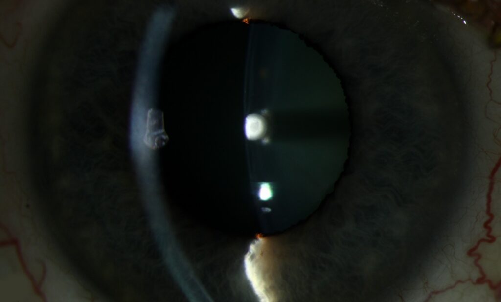
- Small, white, sharply circumscribed opacity at the very front of the lens, visible on direct slit-lamp exam without needing retroillumination
- Typically bilateral, though asymmetric in size between the two eyes
- Surrounding lens generally clear
- No associated capsular thinning or risk features, unlike the posterior form
- A dulled or asymmetric red reflex on the Bruckner test in a larger lesion, which is often what actually prompts referral in an infant too young for formal acuity testing
Differential Diagnosis
- Anterior pyramidal cataract — the more aggressive, protruding variant, distinguished by its cone shape and tendency to progress
- Posterior polar cataract — located at the back rather than front of the lens, with a very different surgical risk profile
- Mittendorf’s dot — a posterior capsule finding from a persistent hyaloid remnant, not an anterior lesion
- Anterior subcapsular cataract (acquired) — typically related to prior trauma or chronic anterior segment inflammation, without the sharply demarcated congenital appearance
- Anterior lenticonus — a conical bulging of the anterior lens surface itself, most classically associated with Alport syndrome, distinguished on careful slit-lamp exam by its curved rather than sharply demarcated opacity and its characteristic “oil droplet” appearance on retroillumination
Management
Small, stable, visually insignificant anterior polar cataracts require no treatment beyond routine monitoring, important in children to confirm the opacity is not interfering with normal visual development, with periodic reassessment of visual acuity, fixation behavior, and refraction as the child grows and becomes able to cooperate with more formal testing.
Larger, visually significant lesions — or the more aggressive pyramidal variant — may need surgical removal, which, unlike posterior polar cataract surgery, does not carry an elevated risk of posterior capsule rupture, because the capsular abnormality here is confined to the front of the lens.
In a young child with a visually significant anterior polar cataract, prompt referral and, where indicated, surgery combined with amblyopia management (patching or optical penalization of the fellow eye as needed) follows the same principles used for any visually significant congenital cataract, with the timing of intervention weighed carefully against the risks of surgery in a very young eye and the developmental cost of leaving a visually meaningful opacity untreated during the critical period.
Because most anterior polar cataracts remain stable rather than progressing, the great majority of affected children and adults ultimately need nothing more than periodic observation, making appropriately distinguishing the small minority who do need surgical referral from the majority who do not the central practical task once the diagnosis is made.


Document what you see
Two smartphone imaging tools built for everyday clinic use — one for the slit lamp, one for the fundus.
From Choroida — the team behind this siteReferences
- Bienfang DC, Kelly ME. Anterior polar cataract. In: Krachmer JH, Mannis MJ, Holland EJ, eds. Cornea and Anterior Segment.
- Merin S. Inherited Eye Diseases: Diagnosis and Management.
- American Academy of Ophthalmology. Basic and Clinical Science Course, Section 6: Pediatric Ophthalmology and Strabismus.
- American Academy of Ophthalmology. Basic and Clinical Science Course, Section 11: Lens and Cataract.
Test yourself
A few questions straight from this article.
-
Where does the opacity of an anterior polar cataract sit?
It is a small, sharply demarcated white opacity lying just under the anterior capsule at the centre of the pupillary axis. -
Which developmental failure produces an anterior polar cataract?
A localized failure of separation between the lens vesicle and overlying surface ectoderm leaves abnormal epithelial or fibrous tissue adherent to the central anterior capsule. -
What size is an anterior polar cataract typically?
The opacity is usually 1 to 3 mm and centred on the visual axis, so visual impact depends heavily on its size. -
How is anterior polar cataract usually distributed between the two eyes?
Anterior polar cataract is typically bilateral though asymmetric between eyes, with the surrounding lens generally clear. -
Which screening finding usually prompts referral of an infant with a larger anterior polar cataract?
In an infant too young for formal acuity testing, a dulled or asymmetric red reflex on the Bruckner test is often what actually triggers referral. -
Which variant of anterior polar cataract warrants closer follow-up and more urgency?
Anterior pyramidal cataract is a cone-shaped opacity that can protrude through the pupil, is more often progressive, and is associated with uveitis-like inflammatory changes. -
Anterior lenticonus with an oil-droplet retroillumination reflex is classically linked to which condition?
Anterior lenticonus is a conical bulging of the anterior lens surface most classically associated with Alport syndrome, with a curved opacity and oil-droplet reflex rather than a sharp plaque. -
How does the surgical risk of anterior polar cataract compare with the posterior polar form?
The capsular abnormality is confined to the front of the lens and the posterior capsule is normal, so surgery lacks the elevated rupture risk of the posterior polar form. -
How is a small, visually insignificant anterior polar cataract managed in a child?
Small stable lesions need no treatment beyond routine monitoring, which in children confirms the opacity is not interfering with normal visual development. -
A patient with a known anterior polar cataract reports fluctuating, worsening vision. What does this suggest?
The opacity does not change day to day, so variable or worsening vision around a known anterior polar cataract should prompt a search for an entirely separate cause.
