
Diabetes and cataract are linked in a way that every ophthalmologist and diabetologist should understand.
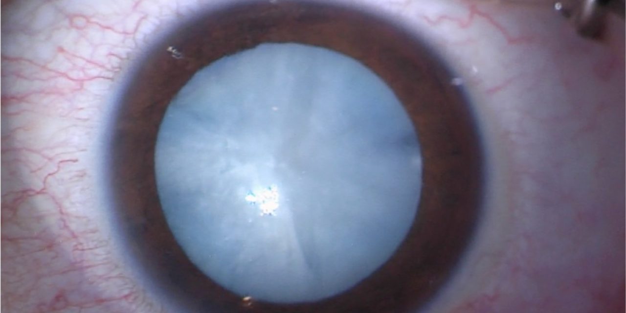
People with diabetes develop cataract at a younger age, progress faster, and face more complications when they have surgery.
Questions about it are among the most frequently searched terms on eye topics, and the answers are practical.
How much does diabetes raise the risk
Population studies, including the Beaver Dam, Blue Mountains, and Wisconsin epidemiologic studies, show that diabetes increases the risk of cataract, with the greatest increase in posterior subcapsular and cortical opacities.
Diabetic patients develop cataract about two to five times more often than non-diabetic patients in younger age groups, and have a 60 percent higher chance of needing cataract surgery overall.
Risk rises with duration of diabetes and poor glycemic control.
Two forms of diabetic cataract
True diabetic (snowflake) cataract
This is uncommon and occurs in young people with type 1 diabetes and very high blood glucose.
Bilateral cortical opacities appear rapidly, over days to weeks, as snowflake-like or flaky white spots, and the lens may become entirely opaque (see snowflake cataract).
The mechanism is osmotic.
High glucose is converted by aldose reductase into sorbitol, which accumulates in the lens fibers and draws in water.
Age-related cataract at a younger age
Much more common.
Nuclear, cortical and posterior subcapsular opacities develop earlier than expected.
Glycation of lens proteins, oxidative stress, and the polyol pathway are all involved.
Fundus Explorer Pro
Photograph the retinal findings described here with the phone already in your pocket — 22 D optics and built-in illumination in one handheld unit.
From Choroida — the team behind this siteWhat patients notice
Blurred or fluctuating vision, glare, and reading difficulty.
Fluctuations in refraction with changes in blood glucose are common: a hyperglycemic myopic shift in the lens, followed by a hyperopic shift when glucose is corrected.
A patient who has newly started treatment may notice vision worsening for a few weeks before it settles, and spectacle prescriptions should not be changed until glucose has been stable for a few weeks.
Preparing for surgery
Cataract surgery in a diabetic patient is routine in most cases, but a few additional steps matter.
- Examine the retina fully before surgery, with dilated fundoscopy and OCT. Cataract can hide diabetic retinopathy, and surgery can accelerate its progression.
- Treat diabetic macular edema before surgery, with anti-VEGF injections if possible, since edema after surgery is more likely and harder to treat (see diabetic macular edema).
- Treat proliferative disease with laser before surgery when the view allows.
- Optimize glycemic control. A recent HbA1c is useful, and an extremely high value should prompt a delay if it is safe to do so.
- Consider topical NSAIDs in addition to corticosteroids to lower the risk of macular edema (see pseudophakic cystoid macular edema).
- Be aware of the higher risk of infection, and consider intracameral antibiotics.
After surgery
Close follow-up is important.
The retina should be examined for progression of diabetic retinopathy, and OCT at four to six weeks can detect macular edema before the patient notices it.
Posterior capsule opacification is also more frequent in diabetic patients (see Elschnig pearls and posterior capsule opacification).
Prevention
Good control of blood glucose, blood pressure, and lipids reduces the risk of cataract, as well as retinopathy.
Stopping smoking and protecting the eyes from ultraviolet light help too.
Questions patients ask
- Will surgery make my retinopathy worse? It can in some eyes, which is why the retina is examined before surgery and treated first when needed.
- Can I have both eyes done on the same day? Most surgeons operate on one eye at a time in diabetic patients, so that macular edema or infection in the first eye can be recognized before the second is treated.
- Will tight sugar control reverse my cataract? It will not clear an established opacity, but it slows the progression of new ones and reduces the risk of surgical complications.
- Do I need to stop my diabetes tablets before surgery? Follow the instructions of the surgical team and the diabetologist, because they depend on the type of anesthesia and the schedule of the day.
Aftercare
Patients should use their drops as prescribed, keep appointments, and monitor their sugar, since high levels slow healing. A review at four to six weeks, with OCT, is a sensible routine to catch macular edema before it affects vision.
Prognosis
Most people with diabetes achieve a good visual result after cataract surgery.
The final outcome depends mainly on the condition of the retina.


Document what you see
Two smartphone imaging tools built for everyday clinic use — one for the slit lamp, one for the fundus.
From Choroida — the team behind this siteReferences
- Klein BE, Klein R, Moss SE. Incidence of cataract surgery in the Wisconsin Epidemiologic Study of Diabetic Retinopathy. Am J Ophthalmol. 1995;119:295-300.
- Rowe NG, Mitchell PG, Cumming RG, Wans JJ. Diabetes, fasting blood glucose and age-related cataract: the Blue Mountains Eye Study. Ophthalmic Epidemiol. 2000;7:103-114.
- Chew EY, Benson WE, Remaley NA, et al. Results after lens extraction in patients with diabetic retinopathy: early treatment diabetic retinopathy study report number 25. Arch Ophthalmol. 1999;117:1600-1606.
- Denniston AK, Chakravarthy U, Zhu H, et al. The UK Diabetic Retinopathy Electronic Medical Record (UK DR EMR) Users Group, Report 2: real-world data for the impact of cataract surgery on diabetic macular oedema. Br J Ophthalmol. 2017;101:1673-1678.
