
Vernal keratoconjunctivitis is the diagnosis that separates itself from ordinary seasonal allergic conjunctivitis by one specific, unmistakable finding: giant, cobblestone-shaped papillae on the upper tarsal conjunctiva, a sign that signals a more severe, chronic allergic process with a genuine capacity to threaten the cornea rather than simply causing itching and redness.
More Than Just Severe Allergies
Vernal keratoconjunctivitis is a chronic, recurrent, severe form of allergic conjunctivitis, typically affecting children and young adults, more common in warm climates, and often with a seasonal pattern, though some cases persist year-round.
What distinguishes it clinically from more common, milder allergic conjunctivitis is both the intensity of the tarsal conjunctival reaction and its recognized potential to involve and damage the cornea, a complication ordinary allergic conjunctivitis essentially never produces.
The Defining Sign: Giant Papillae
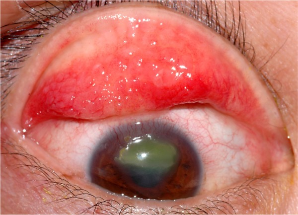
The upper tarsal conjunctiva develops large, flat-topped papillae, often described as having a cobblestone appearance given their size and closely packed arrangement, and this finding, when present, is essentially diagnostic of vernal keratoconjunctivitis rather than a milder allergic process.
- Horner-Trantas dots, small, white, gelatinous limbal nodules composed of degenerated eosinophils, are another characteristic finding, particularly in the limbal variant of this condition
- Limbal involvement can also include limbal thickening and a gelatinous limbal infiltrate, distinct from the tarsal papillary form though both patterns can coexist in the same patient
All-fit Slit-Lamp Adapter
Record and share exactly what you see at the slit lamp. One adapter fits any slit lamp or surgical microscope — and any smartphone.
From Choroida — the team behind this siteWhy the Cornea Is at Risk
The intense inflammatory activity in vernal keratoconjunctivitis, combined with the mechanical effect of giant papillae rubbing against the corneal surface with every blink, can produce a spectrum of corneal complications not seen in milder allergic conditions.
- Superficial punctate keratopathy is common during active disease, related to both direct inflammatory mediator effects and mechanical trauma from the papillae
- In severe, poorly controlled cases, a shield ulcer can develop, a sterile, typically oval, superior corneal epithelial defect with an associated plaque of inflammatory debris, representing the most serious corneal complication of this condition and one that can threaten vision if not promptly recognized and treated
- Persistent or recurrent shield ulcers can lead to corneal scarring and, in severe cases, visually significant astigmatism
Clinical Features
- Intense itching, often the most prominent and distressing symptom, along with tearing, photophobia, and a thick, ropy mucous discharge characteristic of this condition
- Bilateral involvement is typical, though asymmetry between the two eyes is common
- A history of other atopic conditions, including asthma, eczema, or allergic rhinitis, is frequent, though not universal
Diagnostic Evaluation
- Slit-lamp examination of the everted upper eyelid is essential to identify giant papillae, a step sometimes omitted in a more cursory external examination, but necessary to distinguish this condition from milder allergic conjunctivitis
- Careful corneal examination with fluorescein staining to identify any punctate keratopathy or, in severe cases, shield ulcer formation
- A history characterizing symptom severity, seasonality, and any prior episodes, along with assessment for other atopic conditions
Management
Stepwise Medical Therapy
- Topical antihistamine and mast cell stabilizer combination agents form the foundation of maintenance therapy, reducing the underlying allergic inflammatory drive
- Topical corticosteroids are used for active flares and are often necessary to control the more intense inflammation characteristic of this condition, though with careful monitoring given the risks of prolonged corticosteroid use, including elevated intraocular pressure and cataract
- Topical cyclosporine or tacrolimus provides an effective, corticosteroid-sparing option for longer-term control in patients with frequent flares or corticosteroid dependence
Managing Shield Ulcers
Shield ulcers require prompt, more intensive treatment, including topical corticosteroids to control the underlying inflammation and supportive measures to promote epithelial healing, given the risk of corneal scarring if this complication is not adequately addressed.
Environmental and Supportive Measures
Cool compresses, avoidance of known triggers where identifiable, and environmental modification, such as reducing exposure to allergens during peak seasons, provide additional symptomatic benefit alongside pharmacologic treatment.
Pitfalls in treatment
- Prolonged steroids cause glaucoma and cataract, so use pulses and steroid-sparing drugs
- Antihistamines alone will not control severe disease
- Contact lens wear is poorly tolerated during active disease
- A shield ulcer needs prompt treatment to avoid scarring
What to tell parents
Explain that the disease tends to improve in the teenage years, that it is not contagious, and that avoiding eye rubbing and managing triggers helps. Arrange follow-up every few months and check the pressure at each visit.
Prognosis
Vernal keratoconjunctivitis often improves with age, with many patients experiencing significant reduction in disease activity by late adolescence or early adulthood, though the disease course is quite variable between individuals.
With appropriate, sustained management, including prompt recognition and treatment of corneal complications such as shield ulcers, most patients avoid permanent visual impairment, underscoring the value of recognizing the giant papillae that distinguish this condition from more common, milder forms of allergic conjunctivitis early in the disease course.


Document what you see
Two smartphone imaging tools built for everyday clinic use — one for the slit lamp, one for the fundus.
From Choroida — the team behind this siteReferences
- Bonini S, Bonini S, Lambiase A, et al. Vernal keratoconjunctivitis revisited: a case series of 195 patients with long-term follow-up. Ophthalmology. 2000;107:1157-1163.
- Leonardi A. Management of vernal keratoconjunctivitis. Ophthalmol Ther. 2013;2:73-88.
- Bremond-Gignac D, Donadieu J, Leonardi A, et al. Prevalence of vernal keratoconjunctivitis: a rare disease? Br J Ophthalmol. 2008;92:1097-1102.
- Sridhar MS, Gopinathan U, Rao GN. Shield ulcers. Cornea. 2001;20:265-269.
Test yourself
A few questions straight from this article.
-
Which finding distinguishes vernal keratoconjunctivitis from milder allergic conjunctivitis?
Large, flat-topped papillae with a cobblestone appearance on the upper tarsal conjunctiva are essentially diagnostic of vernal keratoconjunctivitis. They mark a more severe, chronic allergic process than ordinary allergic conjunctivitis. -
Which feature of vernal keratoconjunctivitis is essentially absent in ordinary allergic conjunctivitis?
What sets vernal keratoconjunctivitis apart is the intensity of the tarsal reaction and its recognised potential to involve and damage the cornea. Ordinary allergic conjunctivitis essentially never produces that complication. -
What is the typical long-term course of vernal keratoconjunctivitis?
The condition often improves with age, with many patients showing a significant fall in disease activity by late adolescence or early adulthood, though the course varies between individuals. With sustained management most patients avoid permanent visual impairment. -
What are Horner-Trantas dots composed of?
Horner-Trantas dots are small, white, gelatinous limbal nodules made up of degenerated eosinophils. They are characteristic, particularly of the limbal variant of the condition. -
Why does the cornea come under threat in vernal keratoconjunctivitis?
Intense inflammatory activity combines with the mechanical effect of giant papillae rubbing the corneal surface with every blink. Together these produce corneal complications not seen in milder allergic disease, beginning with superficial punctate keratopathy. -
What is the appearance of a shield ulcer in vernal keratoconjunctivitis?
A shield ulcer is a sterile, typically oval, superior corneal epithelial defect with an associated plaque of inflammatory debris. It is the most serious corneal complication of the condition and can threaten vision if not promptly treated. -
What can persistent or recurrent shield ulcers lead to?
Persistent or recurrent shield ulcers can produce corneal scarring and, in severe cases, visually significant astigmatism. This is why prompt, more intensive treatment of a shield ulcer matters. -
Which examination step is essential to diagnose vernal keratoconjunctivitis?
Slit-lamp examination of the everted upper lid is essential to identify giant papillae and is sometimes omitted in a cursory external examination. Fluorescein staining of the cornea is then used to detect keratopathy or a shield ulcer. -
Which agents form the foundation of maintenance therapy in vernal keratoconjunctivitis?
Combination topical antihistamine and mast cell stabiliser agents form the foundation of maintenance therapy, reducing the underlying allergic inflammatory drive. Antihistamines alone will not control severe disease. -
Which treatment offers corticosteroid-sparing control in frequently flaring vernal keratoconjunctivitis?
Topical cyclosporine or tacrolimus provides effective corticosteroid-sparing control for patients with frequent flares or corticosteroid dependence. This matters because prolonged corticosteroid use carries risks of raised intraocular pressure and cataract.

Writes and reviews the clinical articles on Educate. Every post is checked against peer-reviewed sources before it goes live.


