

Fifteen years ago, a diagnosis of wet macular degeneration meant a slow, predictable slide toward legal blindness.
Today the same diagnosis is met with a needle – and, for most patients, a stable retina for years afterward.
Intravitreal injection is the delivery route that made anti-VEGF therapy possible, and it is now one of the most frequently performed procedures in medicine.
The pharmacology gets the attention, but the outcome in any individual patient is decided just as much by the injection itself – the sterile technique, the timing, the choice between fixed dosing and treat-and-extend.
A well-executed intravitreal injection service is what turns a molecule with excellent trial data into a real-world result.
What Is Intravitreal Injection Therapy?
Intravitreal injection is the direct delivery of a drug into the vitreous cavity through the pars plana, bypassing the blood-retinal barrier that limits systemic or topical drugs from reaching the retina in useful concentration.
The agents delivered this way fall into several classes:
- Anti-VEGF agents – ranibizumab, aflibercept, bevacizumab (used off-label), brolucizumab and the bispecific agent faricimab, which additionally blocks angiopoietin-2
- Corticosteroids – dexamethasone and fluocinolone acetonide implants, for inflammatory and refractory oedema
- Complement inhibitors – pegcetacoplan and avacincaptad pegol, for geographic atrophy
- Antimicrobials – for infectious endophthalmitis
The injection technique itself is essentially identical across all of these; what differs is the frequency and the disease being treated.
Indications
Intravitreal therapy has become first-line treatment across a wide range of retinal disease.
- Neovascular age-related macular degeneration – the original and still the highest-volume indication
- Diabetic macular oedema
- Macular oedema following retinal vein occlusion, both branch and central
- Myopic choroidal neovascularisation
- Geographic atrophy, with complement inhibitors aimed at slowing lesion growth rather than restoring vision
- Endophthalmitis, treated with intravitreal antibiotics as an emergency
The sheer breadth of this list is why injection clinics have become one of the largest single workloads in modern ophthalmology.
Fundus Explorer Pro
Photograph the retinal findings described here with the phone already in your pocket — 22 D optics and built-in illumination in one handheld unit.
From Choroida — the team behind this siteAnti-VEGF Pharmacology
Understanding why the drug is injected, rather than given systemically, explains most of the practical decisions around treatment.
- Vascular endothelial growth factor drives the abnormal vessel growth and vascular leakage underlying wet AMD, diabetic macular oedema and venous occlusion
- Anti-VEGF agents bind and neutralise VEGF, reducing leakage and, in many eyes, causing regression of neovascular complexes
- Bevacizumab, ranibizumab and brolucizumab are single-target anti-VEGF-A agents; aflibercept also binds placental growth factor
- Faricimab is a bispecific antibody blocking both VEGF-A and angiopoietin-2, targeting vessel stabilisation as well as VEGF suppression
- Effect duration varies by molecule, which is what allows some newer agents to be dosed less frequently in treat-and-extend regimens
The clinically important point is that these are not cures – they suppress an active disease process, and most patients need sustained treatment to keep the gains they achieve.
Injection Technique
Pre-Injection Preparation
- Topical antisepsis with povidone-iodine applied to the ocular surface and periocular skin immediately before injection – the single intervention with the strongest evidence for reducing endophthalmitis risk
- Topical anaesthesia, and a lid speculum to keep the lashes clear of the injection site
- Marking or estimating the injection site, typically 3.5 to 4 mm posterior to the limbus in a phakic eye and 3.5 mm in a pseudophakic eye
The Injection
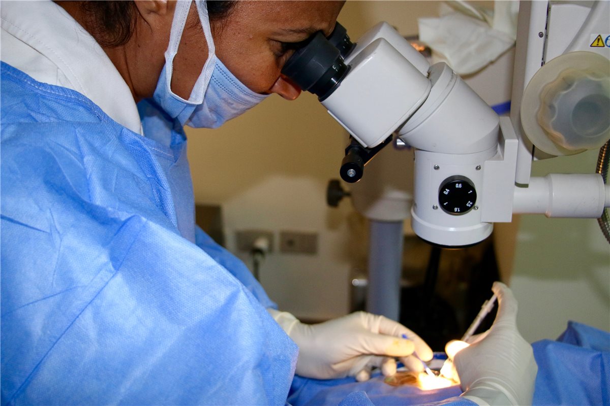
- A fine-gauge needle is passed through the pars plana, aimed toward the mid-vitreous cavity away from the lens and macula
- The drug is delivered as a fixed-volume bolus appropriate to the agent
- The needle is withdrawn and the injection site checked for reflux or a wick of vitreous
Post-Injection Care
- A check of light perception or hand movement vision immediately after injection to exclude central retinal artery occlusion from a pressure spike
- Patient counselling on red-flag symptoms – worsening pain, redness or vision loss – that should prompt urgent same-day review
- No routine post-injection topical antibiotics; evidence does not support their use and they add cost without benefit
Because the whole procedure takes only a few minutes, technique variation between injectors is easy to overlook – yet it is precisely what separates a low-complication clinic from a high one.
Treatment Regimens
Fixed Dosing
- Monthly or bimonthly injections given regardless of disease activity
- Predictable and simple to plan, but the highest treatment burden for patients and clinics
Pro Re Nata (PRN)
- Injection only when disease activity is detected on monthly monitoring
- Reduces injection number but requires frequent visits regardless, and can allow disease to reactivate between assessments
Treat and Extend
- Injections given at every visit, with the interval between visits progressively lengthened when the retina remains dry and shortened if fluid recurs
- Aims to maintain continuous disease control while minimising visit frequency, and has become the dominant real-world strategy
Treat and extend has largely displaced strict PRN in practice because it avoids the monitoring-only visits where disease is found to have already reactivated.
Complications
Injection-Related
- Endophthalmitis – rare but sight-threatening, presenting with pain, redness and vision loss usually within days of injection, requiring emergency vitreous tap and intravitreal antibiotics
- Transient intraocular pressure spike, usually self-limiting but relevant in patients with pre-existing glaucoma
- Subconjunctival haemorrhage – common, cosmetically alarming, but clinically benign
- Retinal detachment or vitreous haemorrhage from inadvertent globe or retinal trauma – rare with correct technique
- Traumatic cataract from lens touch, largely avoidable with correct needle angle and pars plana measurement
Drug-Related
- Sterile intraocular inflammation, reported with some agents more than others, notably brolucizumab
- Rare arterial thromboembolic events with systemic absorption, a theoretical concern that has driven long-term safety monitoring
- Geographic atrophy progression has been reported with long-term anti-VEGF use in some studies, though causation remains debated
Endophthalmitis is the complication that defines injection-day protocol, because outcome depends almost entirely on how quickly it is recognised and treated once it occurs.
Patient Counselling
- Realistic expectation-setting – most patients achieve stabilisation or modest improvement, not full restoration of lost vision
- The long-term nature of treatment, often extending over years, and the impact of appointment burden on adherence
- Clear red-flag symptoms and a direct route to urgent review if they occur
- The rationale for treat-and-extend, since patients who understand why intervals change are more likely to tolerate a shortened interval without alarm
Adherence is, in practice, the single largest determinant of long-term visual outcome across every indication treated this way.
Prognosis
Outcomes depend heavily on the disease being treated and on how consistently therapy is delivered.
- Neovascular AMD treated promptly and consistently maintains vision in the large majority of patients, with meaningful gains in many
- Diabetic macular oedema responds well to anti-VEGF therapy, though a subset of eyes are relatively resistant and may benefit from steroid implants instead
- Delayed or interrupted treatment – common in real-world practice compared with trial conditions – is consistently associated with worse visual outcomes than in clinical trials
- Geographic atrophy treatment slows anatomical progression but has not been shown to improve visual acuity
The gap between trial results and real-world outcomes in this field is almost entirely explained by treatment persistence – the drugs work when they are actually given on schedule.


Document what you see
Two smartphone imaging tools built for everyday clinic use — one for the slit lamp, one for the fundus.
From Choroida — the team behind this siteReferences
- Rosenfeld PJ, Brown DM, Heier JS, et al. Ranibizumab for neovascular age-related macular degeneration. New England Journal of Medicine. 2006.
- Heier JS, Khanani AM, Quezada Ruiz C, et al. Efficacy, durability, and safety of faricimab: TENAYA and LUCERNE trials. The Lancet. 2022.
- Royal College of Ophthalmologists. Intravitreal injection therapy guidelines.
- Anti-VEGF Intravitreal Therapy. EyeWiki, American Academy of Ophthalmology.
- Intravitreal Injections. StatPearls, NCBI Bookshelf.
Test yourself
A few questions straight from this article.
-
What problem does intravitreal injection solve that systemic or topical drug delivery cannot?
Injection through the pars plana delivers drug straight into the vitreous cavity, bypassing the blood-retinal barrier that stops systemic or topical agents reaching the retina in useful concentration. -
Which two targets does the bispecific agent faricimab block?
Faricimab is a bispecific antibody blocking both VEGF-A and angiopoietin-2, so it targets vessel stabilisation as well as VEGF suppression. -
Which pre-injection step has the strongest evidence for reducing endophthalmitis risk?
Topical povidone-iodine applied to the ocular surface and periocular skin immediately before injection is the single intervention with the strongest evidence for reducing endophthalmitis. -
How far posterior to the limbus is the needle passed in a pseudophakic eye?
The injection site is typically 3.5 mm posterior to the limbus in a pseudophakic eye and 3.5 to 4 mm in a phakic eye, keeping the needle within the pars plana. -
Why is vision checked immediately after an intravitreal injection?
A check of light perception or hand movement vision straight after injection excludes central retinal artery occlusion caused by the transient intraocular pressure spike. -
What is the recommended position on topical antibiotics after an intravitreal injection?
Routine post-injection topical antibiotics are not used because the evidence does not support them and they add cost without benefit. -
What defines a treat-and-extend regimen of intravitreal therapy?
Treat and extend injects at every visit and progressively lengthens the interval while the retina remains dry, shortening it if fluid recurs; it has become the dominant real-world strategy. -
How should post-injection endophthalmitis be managed?
Endophthalmitis presents with pain, redness and vision loss usually within days of injection and requires emergency vitreous tap and intravitreal antibiotics, as outcome depends on speed of treatment. -
Sterile intraocular inflammation has been reported notably with which anti-VEGF agent?
Sterile intraocular inflammation is a recognised drug-related complication of intravitreal therapy, reported with some agents more than others and notably with brolucizumab. -
What outcome do complement inhibitors achieve in geographic atrophy?
Complement inhibitors such as pegcetacoplan and avacincaptad pegol aim to slow lesion growth rather than restore vision, and have not been shown to improve visual acuity.

Writes and reviews the clinical articles on Educate. Every post is checked against peer-reviewed sources before it goes live.


