
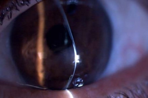
Pellucid marginal degeneration is a peripheral corneal thinning disorder that is often grouped together with keratoconus as a corneal ectasia but has a distinct enough pattern of thinning, topography, and clinical behavior that treating the two as interchangeable leads to poor surgical planning, particularly if crosslinking or refractive surgery is being considered.
The Pattern of Thinning
Unlike keratoconus, in which the area of maximal thinning and steepening typically lies close to the visual axis or paracentrally, pellucid marginal degeneration causes thinning in a narrow band of peripheral inferior cornea, classically located between the four and eight o’clock positions, a short distance from the limbus, while the cornea directly above this band bulges forward and becomes markedly steep.
The central cornea in the visual axis often remains relatively normal in thickness and curvature, which is why some patients with significant pellucid marginal degeneration retain reasonably good corrected vision with glasses or a well-fitted contact lens despite substantial peripheral corneal distortion.
Clinical and Topographic Features
- A thin, crescentic band of inferior peripheral corneal thinning, typically without significant scarring, vascularization, or lipid deposition, distinguishing it from other peripheral corneal thinning disorders such as Terrien marginal degeneration
- High, often irregular, against-the-rule or oblique astigmatism, frequently more severe and harder to correct with glasses than typical keratoconus astigmatism
- The classic “crab-claw” or “butterfly” pattern on corneal topography, showing a bow-tie of steepening below the horizontal meridian with flattening above it, distinct from the more central, asymmetric round or oval steepening typical of keratoconus
- Absence of a Fleischer ring or Vogt striae in the classic, pure form, findings that are common in keratoconus
- Typically bilateral, and onset is often somewhat later than the classic teenage-to-young-adult onset of keratoconus
All-fit Slit-Lamp Adapter
Record and share exactly what you see at the slit lamp. One adapter fits any slit lamp or surgical microscope — and any smartphone.
From Choroida — the team behind this siteWhy the Distinction Matters
- Crosslinking protocols and outcomes data are based primarily on keratoconus, and the peripheral, inferior location of thinning in pellucid marginal degeneration makes standard central crosslinking treatment zones less directly applicable, requiring modified treatment approaches
- Contact lens fitting differs substantially, since the peripheral steepening and relatively normal central cornea in pellucid marginal degeneration often require a different lens design than the central cone typical of keratoconus, and scleral lenses are frequently favored
- Surgical planning for corneal transplantation, when needed, differs because the area of thinning is peripheral and close to the limbus, which affects graft size and shape and increases the technical difficulty and rejection risk compared with a typical central keratoconus graft
- Refractive surgery, such as LASIK, is contraindicated in both conditions, but recognizing pellucid marginal degeneration specifically is important since its topographic pattern can otherwise be under-recognized or misclassified as simple astigmatism on a cursory topography read
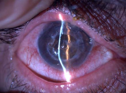
Diagnosis
- Corneal tomography or topography is the key diagnostic tool, showing the characteristic crab-claw pattern
- Pachymetry mapping confirms the peripheral, inferior location of maximal thinning, distinct from the more central thinning pattern of keratoconus
- Slit-lamp examination to assess for the presence or absence of findings such as Fleischer ring, Vogt striae, and corneal scarring, which help support the diagnosis and distinguish it from advanced keratoconus with peripheral extension
Management
- Glasses or, more often given the high irregular astigmatism, rigid gas permeable or scleral contact lenses for optical correction
- Crosslinking, using protocols modified to address the peripheral treatment zone, is used in progressive cases, though the evidence base is less extensive than for classic keratoconus
- Surgical options for advanced disease include peripheral lamellar keratoplasty, crescentic wedge resection, or, in severe cases, full-thickness keratoplasty tailored to the peripheral pattern of involvement, each carrying more technical complexity than standard central keratoconus surgery
Prognosis
Vision can often be well managed with contact lenses for extended periods, and many patients do not require surgical intervention.
When surgery is needed, outcomes are generally less predictable than for central keratoconus surgery, given the peripheral, near-limbal location of the diseased tissue, which is part of why an accurate distinction between the two conditions matters from the earliest stages of workup.


Document what you see
Two smartphone imaging tools built for everyday clinic use — one for the slit lamp, one for the fundus.
From Choroida — the team behind this siteReferences
- Krachmer JH. Pellucid marginal corneal degeneration. Arch Ophthalmol. 1978;96:1217-1221.
- Sridhar MS, Mahesh S, Bansal AK, Nutheti R, Rao GN. Pellucid marginal corneal degeneration. Ophthalmology. 2004;111:1102-1107.
- Rabinowitz YS. Pellucid marginal corneal degeneration. In: Krachmer JH, Mannis MJ, Holland EJ, eds. Cornea. 4th ed. Philadelphia: Elsevier; 2017.
- Kymionis GD, Portaliou DM, Kounis GA, Limnopoulou AN, Kontadakis GA, Grentzelos MA. Corneal collagen cross-linking with riboflavin and UV-A for the treatment of pellucid marginal degeneration. Am J Ophthalmol. 2009;148:70-75.
Test yourself
A few questions straight from this article.
-
Where is the band of thinning in pellucid marginal degeneration?
Pellucid marginal degeneration thins a narrow band of peripheral inferior cornea, classically between the four and eight o'clock positions, a short distance from the limbus. -
Why do some patients with pellucid marginal degeneration keep reasonably good corrected vision?
The cornea in the visual axis often remains relatively normal in thickness and curvature, so good corrected vision is possible with glasses or a well-fitted lens despite marked peripheral distortion. -
Which corneal topography pattern is classic for pellucid marginal degeneration?
The classic crab-claw or butterfly topographic pattern distinguishes pellucid marginal degeneration from the more central, asymmetric round or oval steepening typical of keratoconus. -
Which keratoconus findings are absent in classic pellucid marginal degeneration?
A Fleischer ring and Vogt striae, both common in keratoconus, are absent in the classic pure form of pellucid marginal degeneration. -
Which absent features separate pellucid marginal degeneration from Terrien marginal degeneration?
The crescentic band of thinning in pellucid marginal degeneration typically shows no significant scarring, vascularization or lipid deposition, which separates it from other peripheral thinning disorders such as Terrien marginal degeneration. -
What astigmatism is typical of pellucid marginal degeneration?
Pellucid marginal degeneration produces high, often irregular, against-the-rule or oblique astigmatism that is frequently harder to correct with glasses than typical keratoconus astigmatism. -
Why must crosslinking protocols be modified in pellucid marginal degeneration?
Crosslinking protocols and outcome data come mainly from keratoconus, and the peripheral inferior location of thinning makes standard central treatment zones less applicable, so modified approaches are needed. -
Which contact lens option is frequently favoured in pellucid marginal degeneration?
Because the peripheral steepening and relatively normal central cornea demand a different lens design from a central cone, scleral lenses are frequently favoured. -
Why is corneal transplantation harder in pellucid marginal degeneration than in keratoconus?
The thinning is peripheral and close to the limbus, which affects graft size and shape and increases technical difficulty and rejection risk compared with a typical central keratoconus graft. -
What is the usual long-term course of pellucid marginal degeneration?
Vision can often be well managed with contact lenses for extended periods and many patients never need surgery, while surgical outcomes are less predictable than in central keratoconus.

Writes and reviews the clinical articles on Educate. Every post is checked against peer-reviewed sources before it goes live.


