
CASE REPORT of Plateau Iris
A 69-year-old woman first presented with left eye acute angle-closure with bilateral laser PI done in 1990. She subsequently developed another episode of bilateral acute angle-closure following pupil dilatation despite patent PI in January 2012 and was diagnosed as having Plateau Iris.

Gonioscopy revealed bilateral appositional angle closure with no peripheral anterior synechiae, but no double hump sign was seen. Bilateral ALPI was performed and the attack was subsequently aborted. Since the angles were still narrow, bilateral cataract extraction was performed, right eye in February and left eye in April 2012.
The intraocular pressure (IOP) remained in the range of teens in both eyes with no signs of glaucoma in subsequent follow-up.
In November 2013,
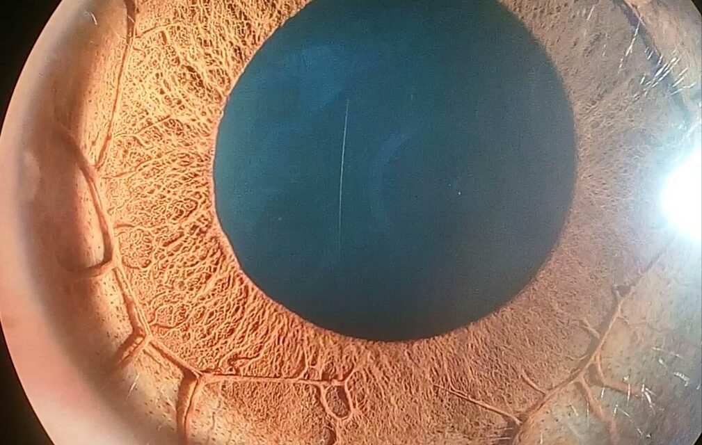
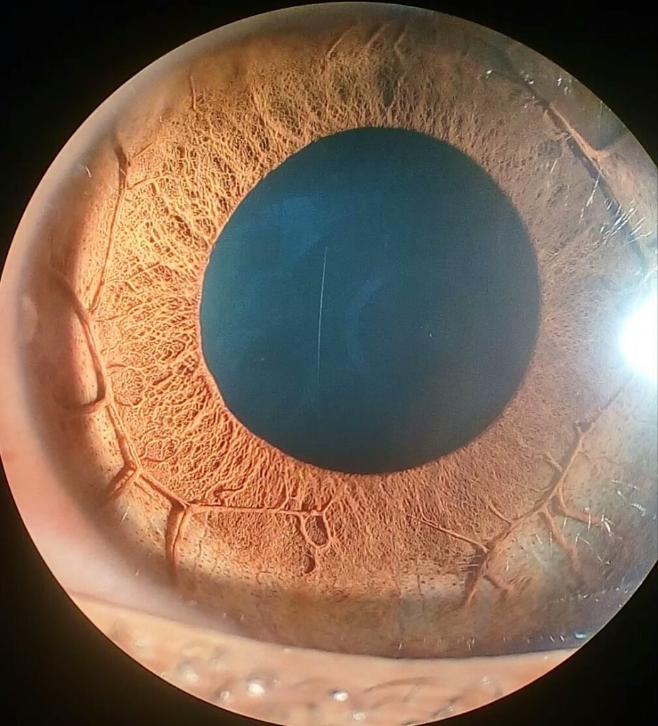
she presented with right eye AAC (IOP 48 mmHg). The cornea was edematous, the anterior chamber was quiet and the central anterior chamber was deep. The PI was patent and the ALPI marks were mostly at peripheral 2/3 of the iris.
The cup-disc ratio was normal. Gonioscopy was suboptimal due to poor corneal clarity but appeared to show grossly narrow angles with patchy closure. The attack was aborted medically. Anterior segment optical coherence tomography confirmed bilateral plateau iris configuration.
DISEASE Plateau Iris
PIS is a type of narrow-angle more commonly seen in younger adults that can lead to chronic angle-closure glaucoma.
Plateau iris refers to the anatomical configuration of the iris. Plateau iris is caused by a narrowing of the anterior chamber angle due to insertion of the iris anteriorly on the ciliary body or displacement of the ciliary body anteriorly, which in turn alters the position of the peripheral iris in relation to the trabecular meshwork (i.e. placing them in apposition).
Plateau iris syndrome is defined as a persistently narrow-angle capable of closure in spite of a patent iridotomy. It is an ocular condition that requires appropriate diagnosis and treatment in order to prevent vision loss.
Early recognition and intervention are key components to a good overall prognosis in this patient population.
MANAGEMENT
The primary treatment modality for patients with plateau iris configuration is surgical. Many clinicians, however, will first treat with miotic agents such as pilocarpine to prevent pupillary dilation leading up to surgery.
Low dose or dilute pilocarpine can produce thinning of the iris and facilitate the opening of the angle by pulling the iris away from the trabecular meshwork.
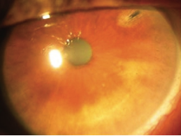
HOW TO TAKE SLIT-LAMP EXAM IMAGES BY A SMARTPHONE?
smartphone slit-lamp photography is the new advancement in the field of science and technology in which the photographs of the desired slit-lamp finding can be taken with smartphones by using the slit-lamp adaptors and Slit lamp smartphone photography.

REFERENCES
- Diniz Filho A, Cronemberger S, Merula RV, Calixto N. Plateau Iris. Arq Bras Oftalmol 2008;71(5):752-8.
- Wand M, Grant WM, Simmons RJ, Hutchinson BT. Plateau Iris Syndrome. Trans Sect Ophthalmol Am Acad Ophthalmol Otolaryngol. 1977;83(1):122-30.
- Ritch R, Chang B, Liebmann J. Angle Closure in Younger Patients. Ophthalmology. 2003; 110(10):1880-9. Review.
- Ultrasound Biomicroscopy (UBM) Clinical Database. Retrieved February 19, 2012.
- Kumar RS, Baskaran M, Chew PT, Friedman DS, Handa S, Lavanya R, et al. Prevalence of plateau iris in primary angle-closure suspects an ultrasound biomicroscopy study. Ophthalmology. Mar 2008;115(3):430-4
Read more about: Tadpole Pupil,Slit lamp Exam, Retinal imaging, Slit lamp exam
