
CASE REPORT
A 53-year-old man presented to an ophthalmology clinic with a complaint of progressive blurring of vision in the right eye for 6 months of duration. The patient claimed that his vision in both eyes was satisfactory prior to this time period.

There were no documented systemic illnesses. His family history was unremarkable. On examination, uncorrected visual acuity was hand movements in the right eye and 6/24 in the left eye.
Refraction was −2.25 −1.50 in the right eye, with no improvement in visual acuity, and +1.75 −2.50 in the left eye, giving a best-corrected visual acuity of 6/6. The intraocular pressure was 14mm Hg in the right eye and 16 mm Hg in the left eye.
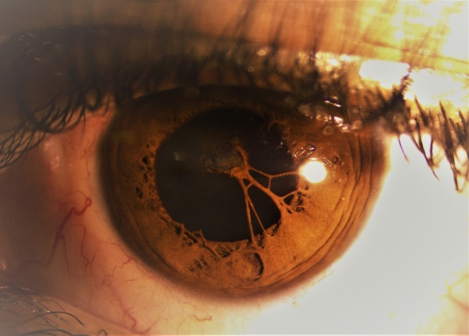
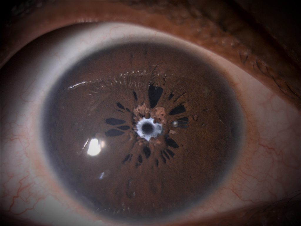
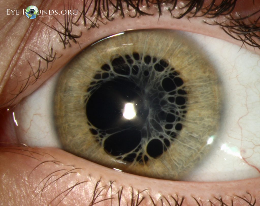
On slitlamp examination, both eyes had clear corneas, deep and quiet anterior chambers, and no synechiae. Both eyes showed a dense network of tissue, more gross and exuberant in the right eye, running from the iris surface and spreading over the pupils.
There was no view of the fundus in the right eye, while the left eye fundus did not reveal any abnormality. B-scan ultrasonography of the right eye revealed a flat retina.
A cover-uncover test was normal. In cases such as this one, tests of macular function like entoptic tests, retina acuity meter (RAM) or laser interferometry can be performed to confirm the status of the macula and the visual prognosis.
The patient was diagnosed with bilateral persistent pupillary membranes (PPM) and cataracts in the right eye.
Persistent Pupillary Membrane DISEASE entity
Persistent pupillary membrane(PPM) is a frequently encountered congenital anomaly. It represents remnants of anterior tunica vasculosa lentis and appears as strands of connective tissue bridging the pupillary area.
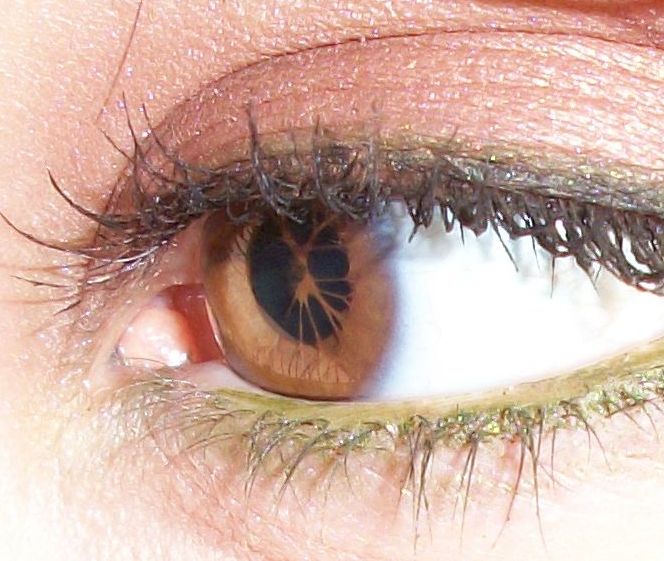
They are usually asymptomatic and of no functional significance. In rare cases, dense membranes can persist and obscure the pupil, causing amblyopia.
Differential Diagnosis
- Post-inflammatory synechie
- Accessory Iris membrane (AIM)- also called iris duplication, closely resembles the normal iris tissue in color and thickness and presents a virtual second pseudo pupil aperture in the center.
- Congenital idiopathic microcoria
- Axenfeld–Rieger syndrome
- Peters anomaly
- Iridocorneal endothelium syndrome (ICE)
Persistent Pupillary Membrane MANAGEMENT
During the first year of life, most PPMs undergo atrophy and require no treatment. Membranes persisting after one year are less likely to regress spontaneously, increasing the risk of deprivational amblyopia. A 1.5-mm pupillary opening is necessary for adequate retinal stimulation and visual cortex development.
Management of Persistent Pupillary Membrane (PPM) depends on the extent of the membrane, and consequently the size of the pupillary opening. Small PPMs can be managed conservatively. Mydriatics, refractive correction, and patching for amblyopia have been used successfully in such cases.
Thick, fibrotic membranes may require surgical excision. Surgery is generally performed in the first weeks or months of life, with a good visual prognosis. It consists of the excision of the pupillary membrane using Vannas or vitreous scissors.
However, surgical management is fraught with risks of anesthesia, intraoperative bleeding, intraocular infection, and cataract formation.
Older patients with thin, sparse membranes may be candidates for Nd: YAG laser membrane lysis. Photodisruption of the membrane can rarely lead to hyphaema. The procedure also carries the risk of cataract formation, iritis, and pigment dispersion.
HOW TO TAKE SLIT-LAMP EXAM IMAGES WITH A SMARTPHONE?
Smartphone slit-lamp photography is the new advancement in the field of science and technology in which photographs of the desired slit-lamp finding can be taken with smartphones by using the slit-lamp adapters.
Slit-lamp Smartphone photography
REFERENCES
- Cassady JR, Light A. Familial Persistent Pupillary Membranes. AMA Arch Ophthalmol. 1957;58(3):438–448.
- Goldberg MF. Persistent fetal vasculature (PVF): An integrated interpretation of signs and symptoms associated with the persistent hyperplastic primary vitreous. Am J Ophthalmol. 1997;124:87–626
- Hadi HA, Hobbs CL. Effect of chronic intrauterine stress on the disappearance of tunica vasculosa lentis of the fetal eye: A neonatal observation Am J Perinatol. 1990;7:23–5
- Tasman W, Jaeger E Duane’s Ophthalmology. 2007 Philadelphia Lippincott Williams & Wilkins:24–258
- Miller SD, Judisch GF. Persistent pupillary membrane: successful medical management. Arch Ophthalmol. 1979 Oct;97(10):1911-3.
- Banigallapati, Shashidhar; Potti, Sudhakar; Marthala, Himabindu A rare case of persistent pupillary membrane, Indian Journal of Ophthalmology: October 2018 – Volume 66 – Issue 10 – p 1480-1483.
