
Retinal macrovessel is a rare congenital vascular anomaly in which an unusually large, aberrant retinal vessel crosses the horizontal raphe, the normal anatomic boundary that otherwise separates the superior and inferior retinal circulations.
It is almost always an incidental finding on routine fundus exam, discovered in an otherwise asymptomatic eye, and its main clinical importance lies in recognizing it correctly rather than mistaking it for something more concerning.
Because it can look visually dramatic on a fundus photograph, a macrovessel unfamiliar to the examiner is easy to over-interpret as pathologic when, in the great majority of cases, it is a benign anatomic variant.
The finding is genuinely rare, and most ophthalmologists will encounter it only occasionally, which is part of why it is easy to misidentify on first exposure without a specific frame of reference for what a benign macrovessel should look like.
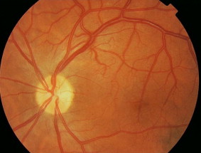
Anatomy and Mechanism
The normal retinal vasculature respects the horizontal raphe as a watershed boundary, with superior and inferior circulations supplied separately and not normally crossing this midline division.
A retinal macrovessel is typically an aberrant retinal artery, occasionally a vein, that develops with an anomalously large caliber and crosses this boundary, most often in the papillomacular or peripapillary region, connecting territory that would normally be served by separate vascular arcades.
The anomaly is congenital, present from birth, and thought to reflect an aberration in normal retinal vascular development rather than an acquired process, though the precise developmental mechanism is not fully established.
Clinical Presentation
- An incidental finding on routine dilated fundus exam in an asymptomatic patient, the most common presentation by far
- A visibly enlarged, tortuous vessel crossing the horizontal raphe, sometimes extending toward or through the macula
- Generally normal visual acuity, since the vessel itself does not typically compromise retinal function despite its striking appearance
- Occasional association with macular edema or exudation when the macrovessel is unusually close to or directly involves the fovea
Fundus Explorer Pro
Photograph the retinal findings described here with the phone already in your pocket — 22 D optics and built-in illumination in one handheld unit.
From Choroida — the team behind this siteAssociated Findings and Complications
Most retinal macrovessels are entirely asymptomatic and require no treatment, behaving as a stable anatomic variant throughout the patient’s life without progression.
A minority of cases are associated with cystoid macular edema, retinal hemorrhage, or exudation, particularly when the anomalous vessel courses directly through or very near the fovea, and these associated findings, not the macrovessel itself, are what actually drive any need for treatment.
Rare reports describe an association between retinal macrovessels and other systemic or ocular anomalies, though the great majority of cases occur in isolation without any other identified abnormality.
Vision loss, when it does occur, is almost always attributable to one of these secondary complications rather than to the presence of the large vessel itself, which is an important distinction to make clear to an understandably concerned patient.
Differential Diagnosis
Retinal arteriovenous malformation, part of the Wyburn-Mason syndrome spectrum discussed in its own dedicated article on this site, involves a more complex tangle of abnormal vessels with direct arteriovenous shunting, distinct from the single large, otherwise normally connected vessel typical of a macrovessel.
Retinal telangiectasia and other vascular anomalies produce a different pattern of abnormal, dilated, or leaking small vessels rather than a single enlarged major vessel crossing the raphe.
A careful fundus exam and, when needed, fluorescein angiography readily distinguish a macrovessel, which shows normal, non-shunting flow through a single large but otherwise unremarkable vessel, from these other, genuinely pathologic vascular anomalies.
Evaluation and Management
Fluorescein angiography confirms the diagnosis by demonstrating normal, laminar blood flow through the enlarged vessel without arteriovenous shunting or leakage, distinguishing a benign macrovessel from a true vascular malformation.
Optical coherence tomography is useful when the macrovessel is near the fovea, to assess for macular edema or structural change that might warrant treatment even though the vessel itself does not.
No treatment is needed for an asymptomatic macrovessel found incidentally with normal vision, and management is reserved specifically for the rare cases with associated macular edema or exudation, following standard treatment approaches for those specific complications.
Periodic monitoring is reasonable given the rarity of the condition and the limited long-term natural history data available, even though most cases remain stable indefinitely without ever requiring intervention.
Patient reassurance matters as much as any specific test here, since an incidentally discovered macrovessel can otherwise cause considerable anxiety for a patient who sees an unusual, striking finding described on their chart or in imaging.


Document what you see
Two smartphone imaging tools built for everyday clinic use — one for the slit lamp, one for the fundus.
From Choroida — the team behind this siteReferences
- Brown GC, Donoso LA, Magargal LE, et al. Congenital retinal macrovessels. Archives of Ophthalmology.
- American Academy of Ophthalmology. Basic and Clinical Science Course, Section 12: Retina and Vitreous.
- Petrella RJ, Wang F, Yannuzzi LA. Congenital retinal macrovessel: report of a case and review of the literature. British Journal of Ophthalmology.
