
Case Study
A 52-year-old woman presented to the emergency clinic with a sudden onset of floaters and flashes of light in her right eye.

Over the past two days, she had also noticed a shadow encroaching from her peripheral vision. Despite these symptoms, her central vision remained unaffected.
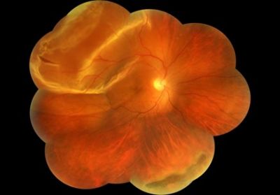
Her visual acuity was 20/20 in the affected eye. Fundoscopic examination revealed a superior retinal detachment sparing the macula and an identifiable retinal tear.
Urgent surgical intervention preserved her macular function, and her visual acuity remained intact postoperatively.
Disease Entity
Macula-on rhegmatogenous retinal detachment (RRD) refers to a specific type of retinal detachment in which the central retina, the macula, remains attached while other parts of the retina are detached.
This condition is an ophthalmic emergency, as prompt treatment can prevent macular involvement and preserve central vision.
Rhegmatogenous retinal detachment arises from a retinal tear or break, allowing vitreous fluid to seep underneath and separate the retina from the underlying layers. In the macula-on variant, detachment is limited to the peripheral retina, maintaining the macula’s function and providing a critical window for intervention.
All-fit Slit-Lamp Adapter
Record and share exactly what you see at the slit lamp. One adapter fits any slit lamp or surgical microscope — and any smartphone.
From Choroida — the team behind this sitePathophysiology
The pathogenesis of macula-on RRD involves three primary factors:
- Retinal Break Formation: Retinal tears, often caused by posterior vitreous detachment (PVD), create a gateway for subretinal fluid to accumulate.
- Subretinal Fluid Accumulation: Fluid infiltrates the subretinal space, causing localized detachment.
- Tractional Forces: Vitreoretinal adhesions exert traction on the retina, exacerbating the detachment.
Sparing of the macula is primarily due to the anatomical location of the retinal tear and the timing of fluid accumulation.
Epidemiology
Macula-on RRD is a relatively rare subtype of retinal detachment but warrants special attention due to its favorable visual prognosis if treated promptly.
- Incidence: Rhegmatogenous retinal detachment has an overall incidence of approximately 10–18 per 100,000 individuals annually, with macula-on cases representing a subset.
- Risk Factors:
- Age: Most common in individuals aged 50–70 years.
- Myopia: Higher risk due to elongated axial length and thinner retina.
- Trauma: Direct retinal injury can precipitate tears.
- History of retinal detachment in the fellow eye.
Clinical Features
Macula-on RRD presents with hallmark symptoms and findings.
- Symptoms:
- Sudden onset of floaters or flashes of light.
- Peripheral visual field loss is described as a shadow or curtain.
- Intact central vision unless progression occurs.
- Fundoscopic Findings:
- Retinal detachment with a distinct area of elevation.
- Retinal breaks or tears are often visible.
- Sparing of the macular area, evident on slit-lamp biomicroscopy.
- Imaging Features:
- Optical Coherence Tomography (OCT): Demonstrates preserved macular architecture.
- Ultrasound B-scan: Useful in cases of media opacities, confirming the extent of detachment.
Diagnosis
The diagnosis of macula-on RRD is primarily clinical, supported by imaging.
- History and Symptoms: Patients often report floaters, flashes, and peripheral vision loss.
- Ophthalmic Examination: Comprehensive retinal evaluation identifies detachment and any associated breaks.
- Imaging:
- OCT to confirm macular integrity.
- Fundus photography and fluorescein angiography may aid in surgical planning.
Differential Diagnosis
Macula-on RRD must be differentiated from other retinal and vitreoretinal conditions.
- Macula-off RRD: Involves macular detachment, leading to central vision loss.
- Vitreous Hemorrhage: Presents with floaters but without detachment.
- Retinoschisis: Mimics detachment but lacks retinal breaks.
- Posterior Vitreous Detachment (PVD): Often a precursor without detachment.
Management
Timely surgical intervention is the cornerstone of macula-on RRD management.
- Preoperative Care:
- Patient education on the importance of head positioning to minimize macular involvement.
- Close monitoring in cases where surgery is delayed.
- Surgical Options:
- Scleral Buckling: Ideal for uncomplicated cases with peripheral tears.
- Pars Plana Vitrectomy (PPV): Suitable for complex tears or significant traction.
- Pneumatic Retinopexy: In selected cases with superior breaks, involving gas bubble tamponade.
- Postoperative Care:
- Positioning instructions, such as face-down positioning after gas tamponade.
- Monitoring for complications like recurrent detachment or proliferative vitreoretinopathy (PVR).
Prognosis
The prognosis for macula-on RRD is excellent with prompt intervention.
- Visual Outcomes: Most patients retain or regain their pre-detachment visual acuity.
- Recurrence Risk: Approximately 5–15%, depending on the presence of PVR or inadequate tear closure.
Prevention
Preventive measures focus on reducing risk factors and early detection.
- Routine Eye Examinations: For high-risk groups, such as myopes or individuals with a history of detachment.
- Patient Education: Awareness of warning signs like floaters and flashes.
- Prophylactic Treatment: Laser photocoagulation or cryotherapy for high-risk retinal tears.
Conclusion
Macula-on rhegmatogenous retinal detachment represents an ophthalmic emergency with a critical opportunity for vision preservation.
Early diagnosis and surgical intervention are paramount in preventing macular involvement and maintaining excellent visual outcomes.
Advances in imaging and surgical techniques have significantly enhanced the management of this condition, ensuring better prognosis and quality of life for affected patients.


Document what you see
Two smartphone imaging tools built for everyday clinic use — one for the slit lamp, one for the fundus.
From Choroida — the team behind this siteReferences
- Goezinne F, La Heij EC, Berendschot TT, et al. Visual outcomes after macula-on versus macula-off retinal detachment surgery. American Journal of Ophthalmology. 2008;146(3):456-463.
- Schwartz SG, Flynn HW Jr. Primary retinal detachment: Clinical features, surgical management, and long-term outcomes. Ophthalmology. 2020;127(9):1208-1213.
- Sodhi A, Leske DA, Campbell JP. Advances in the diagnosis and management of retinal detachment. Retina. 2019;39(4):737-748.
- Chang S. Pneumatic retinopexy: Indications and outcomes. Survey of Ophthalmology. 2008;53(4):443-456.
- Mitry D, Chalmers J, Anderson K, et al. Temporal trends in retinal detachment incidence and management. Ophthalmology. 2011;118(4):795-801.
Test yourself
A few questions straight from this article.
-
What defines a macula-on rhegmatogenous retinal detachment?
In the macula-on variant, detachment is limited to the peripheral retina and the macula stays attached, preserving central vision and creating a window for intervention. -
Which event most commonly initiates the retinal break in rhegmatogenous retinal detachment?
Retinal tears are often caused by posterior vitreous detachment, which creates a gateway for vitreous fluid to accumulate in the subretinal space. -
What is the reported annual incidence of rhegmatogenous retinal detachment?
The article gives an overall incidence of approximately 10 to 18 per 100,000 individuals annually, with macula-on cases forming a subset. -
In which age group is rhegmatogenous retinal detachment most common?
Age is a listed risk factor, with rhegmatogenous retinal detachment most common in individuals aged 50 to 70 years. -
Which symptom triad typically heralds a macula-on rhegmatogenous retinal detachment?
Patients report sudden floaters or flashes and peripheral field loss described as a shadow or curtain, while central vision stays intact unless the detachment progresses. -
Which imaging test confirms that the macula is still attached in rhegmatogenous retinal detachment?
OCT demonstrates preserved macular architecture and is used to confirm macular integrity; ultrasound B-scan helps when media opacities obscure the view. -
Which condition mimics retinal detachment but characteristically lacks retinal breaks?
The article lists retinoschisis as a mimic of detachment that lacks retinal breaks, distinguishing it from rhegmatogenous retinal detachment. -
Which surgical option suits an uncomplicated macula-on detachment with peripheral tears?
Scleral buckling is described as ideal for uncomplicated cases with peripheral tears, while vitrectomy suits complex tears or significant traction. -
Pneumatic retinopexy is best suited to macula-on detachments with breaks in which location?
Pneumatic retinopexy with gas bubble tamponade is used in selected cases with superior breaks. -
What is the approximate recurrence risk after repair of rhegmatogenous retinal detachment?
Recurrence risk is roughly 5 to 15%, depending on proliferative vitreoretinopathy or inadequate closure of the retinal tear.
