
Introduction
Retinal detachment (RD) is an ophthalmic emergency requiring prompt diagnosis and treatment to prevent vision loss.
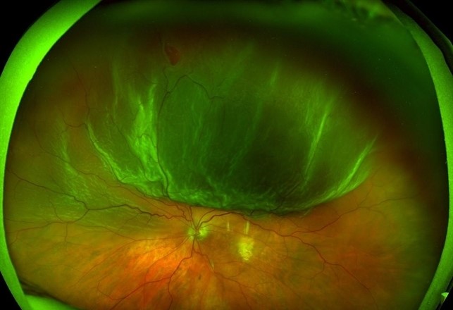
However, several conditions can mimic its presentation, leading to potential misdiagnoses. Differentiating true retinal detachment from its lookalikes is crucial for appropriate management.
This article explores conditions that can resemble RD, highlighting key clinical and imaging findings to distinguish them.
Understanding Retinal Detachment
Retinal detachment occurs when the neurosensory retina separates from the underlying retinal pigment epithelium (RPE). The three main types of RD include:
- Rhegmatogenous RD (RRD): Caused by a retinal break, allowing vitreous fluid to enter and separate the retina.
- Tractional RD (TRD): Due to fibrovascular proliferation pulling the retina away, it is common in diabetic retinopathy.
- Exudative (Serous) RD: Occurs from fluid accumulation beneath the retina without a retinal break, often due to inflammatory, neoplastic, or vascular causes.
Fundus Explorer Pro
Photograph the retinal findings described here with the phone already in your pocket — 22 D optics and built-in illumination in one handheld unit.
From Choroida — the team behind this siteConditions That Mimic Retinal Detachment
1. Vitreous Detachment
Key Features:
- Posterior vitreous detachment (PVD) is a common age-related condition where the vitreous separates from the retina.
- Symptoms include flashes, floaters, and reduced contrast sensitivity, similar to RRD.
- Unlike RD, no retinal elevation or subretinal fluid is present.
- B-scan ultrasound can confirm PVD if the posterior hyaloid face is visible but mobile.

2. Retinoschisis
Key Features:
- A splitting of the retinal layers, often in the inferotemporal quadrant.
- No retinal breaks or vitreous pigment cells.
- Smooth, convex elevation of the retina with an “immobile” appearance on B-scan.
- Optical coherence tomography (OCT) shows a characteristic “schisis cavity.”
3. Choroidal Detachment
Key Features:
- Often caused by hypotony, inflammation, or trauma.
- Presents as smooth, dome-shaped elevations of the choroid.
- Unlike RD, choroidal detachments are more peripheral, firm, and do not shift with eye movement.
- B-scan ultrasound shows suprachoroidal fluid without the typical “swaying” motion of RD.
4. Central Serous Chorioretinopathy (CSCR)
Key Features:
- Serous detachment of the macula due to choroidal hyperpermeability.
- More localized compared to exudative RD.
- No retinal tears or breaks.
- Fluorescein angiography shows “smokestack” or “inkblot” leakage patterns.
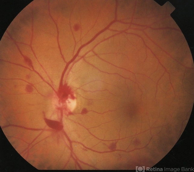
5. Intraocular Tumors (e.g., Choroidal Melanoma, Metastases, Hemangiomas)
Key Features:
- Tumors can elevate the retina, simulating an RD.
- Differentiated by an associated mass, often pigmented.
- B-scan ultrasound helps distinguish solid tumors from fluid-filled RD.
- Optical coherence tomography (OCT) and fluorescein angiography provide additional confirmation.
6. Uveal Effusion Syndrome
Key Features:
- Spontaneous choroidal effusion leading to serous RD.
- No evidence of inflammation or mass.
- Often associated with nanophthalmos or scleral abnormalities.
- OCT and ultrasound imaging confirm suprachoroidal fluid.
7. Inflammatory and Infectious Conditions
Vogt-Koyanagi-Harada Disease (VKH)
- Bilateral serous retinal detachments.
- Associated with systemic autoimmune symptoms.
- Fluorescein angiography shows “hot” choroidal leakage points.
Endophthalmitis or Posterior Uveitis
- Inflammatory exudates mimic RD but are associated with vitritis.
- B-scan ultrasound may show dense vitreous opacities.
- A history of infection or recent intraocular surgery aids in diagnosis.
Diagnostic Approach
Clinical Examination
- Detailed history, including onset, symptoms, and risk factors.
- Dilated fundus examination with indirect ophthalmoscopy.
- The presence of Shaffer’s sign (pigment cells in the vitreous) suggests RRD.
Imaging Modalities
- Optical Coherence Tomography (OCT) – Differentiates between schisis, CSCR, and RD.
- B-Scan Ultrasound – Helps in detecting RD in cases with media opacity.
- Fluorescein Angiography – Useful in inflammatory and vascular conditions.
- Indocyanine Green Angiography – Evaluates choroidal pathology.
Conclusion
Retinal detachment is a vision-threatening condition requiring urgent intervention, but several lookalike conditions must be considered to ensure accurate diagnosis and appropriate treatment.
A thorough clinical examination combined with imaging techniques can help differentiate RD from its mimics, preventing unnecessary surgical interventions and optimizing patient care.


Document what you see
Two smartphone imaging tools built for everyday clinic use — one for the slit lamp, one for the fundus.
From Choroida — the team behind this siteReferences
- Gass JDM. Stereoscopic Atlas of Macular Diseases: Diagnosis and Treatment. Mosby; 1997.
- Yannuzzi LA, Negrão S, Iida T, et al. Retinal angiography and optical coherence tomography findings in central serous chorioretinopathy. Retina. 2003;23(6):769-777.
- Shields CL, Kaliki S, Furuta M, et al. Clinical spectrum and prognosis of choroidal melanocytic tumors in 8036 consecutive eyes. Ophthalmology. 2012;119(1):50-59.
- Johnson MW. Posterior Vitreous Detachment: Evolution and Complications of Its Early Stages. Am J Ophthalmol. 2010;149(3):371-382.
- Spaide RF, Yannuzzi LA, Slakter JS, et al. Indocyanine green angiography in uveal effusion syndrome. Ophthalmology. 1996;103(5):256-262.
Test yourself
A few questions straight from this article.
-
Retinal detachment is defined by separation of the neurosensory retina from which layer?
Retinal detachment occurs when the neurosensory retina separates from the underlying retinal pigment epithelium. -
Which mechanism defines rhegmatogenous retinal detachment?
In rhegmatogenous detachment a retinal break lets vitreous fluid enter and separate the retina, unlike tractional or exudative types. -
Which type of retinal detachment is most characteristic of advanced diabetic retinopathy?
Tractional detachment results from fibrovascular proliferation pulling the retina away and is common in diabetic retinopathy. -
Which feature separates posterior vitreous detachment from rhegmatogenous retinal detachment despite shared flashes and floaters?
PVD produces flashes, floaters, and reduced contrast sensitivity but no retinal elevation or subretinal fluid; B-scan shows a visible but mobile posterior hyaloid. -
Which B-scan and OCT findings support retinoschisis rather than retinal detachment?
Retinoschisis splits the retinal layers, typically inferotemporally, giving a smooth convex immobile elevation and a characteristic schisis cavity on OCT. -
Which features distinguish choroidal detachment from retinal detachment on examination and ultrasound?
Choroidal detachments follow hypotony, inflammation, or trauma and appear as smooth peripheral dome-shaped firm elevations that do not sway on B-scan. -
Which fluorescein angiography patterns are characteristic of central serous chorioretinopathy?
CSCR is a localized serous macular detachment from choroidal hyperpermeability, with no retinal break and smokestack or inkblot leakage on angiography. -
Which condition causes spontaneous choroidal effusion with serous detachment and is linked to nanophthalmos?
Uveal effusion syndrome shows suprachoroidal fluid on ultrasound and OCT without inflammation or mass, often with nanophthalmos or scleral abnormalities. -
Which presentation suggests Vogt-Koyanagi-Harada disease rather than rhegmatogenous retinal detachment?
VKH causes bilateral serous retinal detachments with systemic autoimmune features and hot choroidal leakage points on fluorescein angiography. -
Which clinical sign on examination points to rhegmatogenous retinal detachment?
Shaffer's sign, pigment cells in the vitreous seen on dilated indirect ophthalmoscopy, suggests a rhegmatogenous detachment.
