
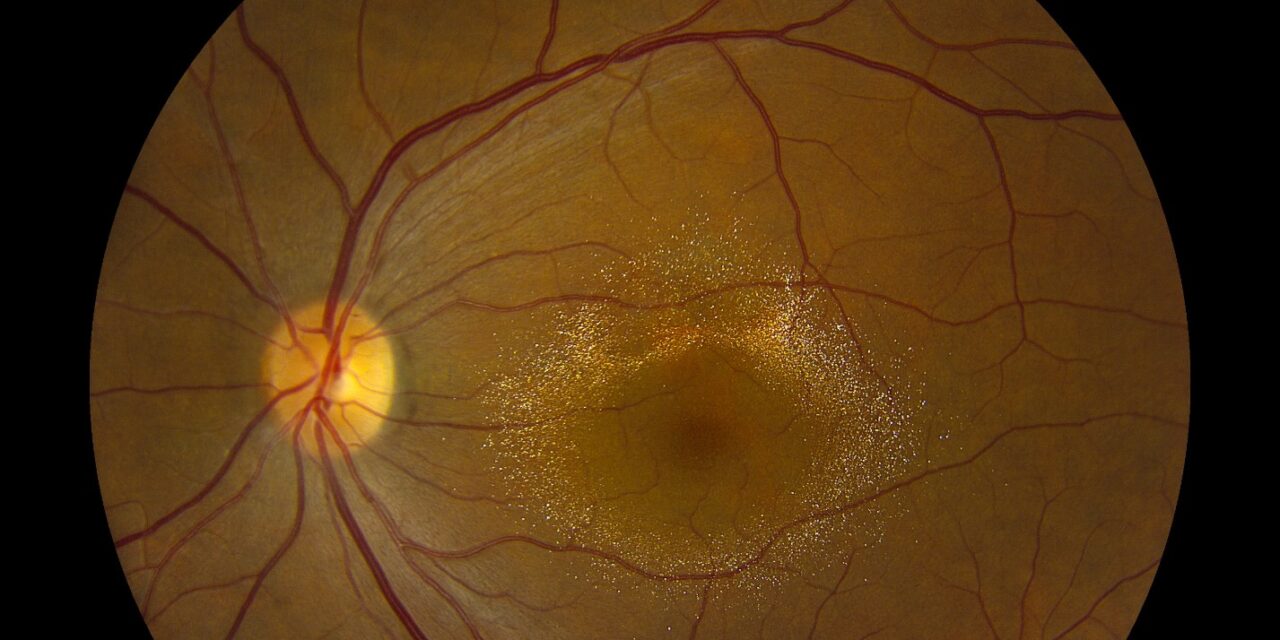
Canthaxanthin retinopathy is a distinctive, dose-related crystalline maculopathy caused by accumulation of canthaxanthin — a carotenoid pigment historically used as an oral “tanning pill” for cosmetic skin bronzing and, separately, as a food coloring and poultry feed additive — within the retina, producing a striking, easily recognized ring of glistening golden-yellow crystals around the fovea.
The finding is a good illustration of how a substance intended purely for a cosmetic or dietary purpose, generally considered “natural” or benign by the people using it, can produce a genuine, objectively visible retinal deposition disorder, even though the functional consequences turn out to be reassuringly mild in the great majority of cases.
Source and Mechanism
Canthaxanthin, taken orally over an extended period — historically most often as a tanning supplement, though also relevant with heavy, prolonged consumption of foods or supplements containing the compound as a coloring agent — is absorbed and distributed throughout the body’s lipid-containing tissues, including the retina, where it deposits in a distinctive pattern within the inner retinal layers surrounding the fovea.
The degree of deposition correlates with cumulative dose, meaning the retinal finding is essentially a visible marker of total lifetime canthaxanthin exposure rather than developing from a single or short course of use.
This dose-dependence is why the finding is more consistently seen in individuals who used tanning supplements regularly over months to years than in those with only brief or occasional exposure, and why a careful history of duration and frequency of use, not simply whether the substance was ever taken, is the most useful piece of exposure history to elicit.
Clinical Presentation
The overwhelming majority of patients with canthaxanthin retinopathy are visually asymptomatic, even with a fully developed, visually striking crystalline ring on fundus exam — this dissociation between a dramatic-appearing fundus finding and essentially normal visual function is one of the most reassuring and clinically important features of the condition, distinguishing it from many other crystalline or deposition maculopathies that do carry a more significant functional risk.
The finding is typically discovered incidentally during a routine dilated fundus exam rather than because of any visual complaint prompting the visit, sometimes surprising a patient who had not disclosed their supplement use because they did not think it was medically relevant.
Fundus Explorer Pro
Photograph the retinal findings described here with the phone already in your pocket — 22 D optics and built-in illumination in one handheld unit.
From Choroida — the team behind this siteExam Findings
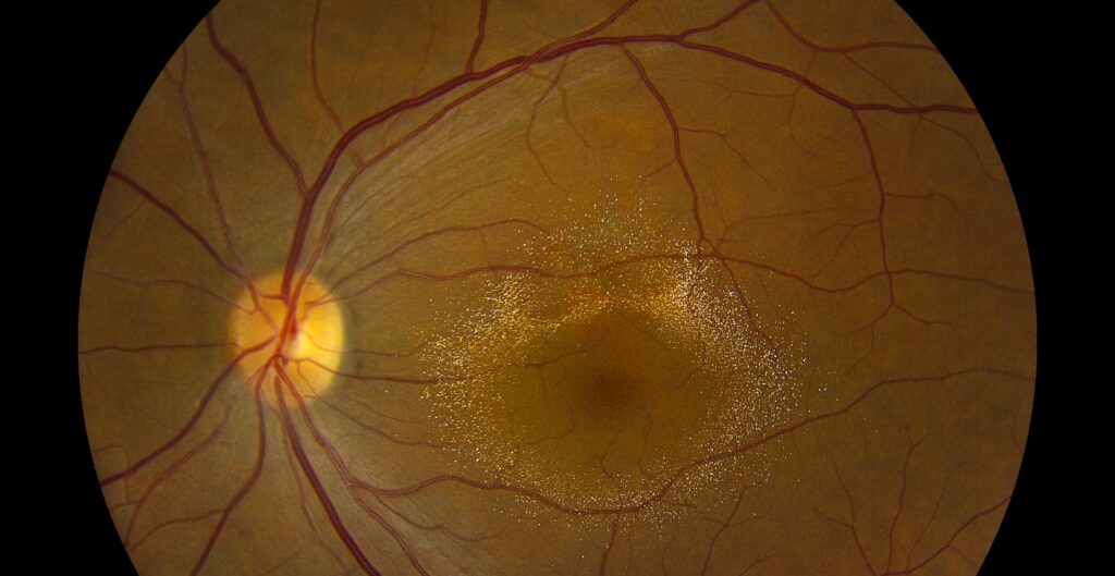
- A ring of fine, glistening, golden-yellow to orange crystals concentrated in the perifoveal region, typically symmetric and bilateral, forming a genuinely distinctive and easily recognized pattern once seen
- Normal visual acuity and normal central visual function in the great majority of cases, even with dense, well-developed crystal deposition
- Generally normal electroretinogram and Amsler grid testing, reflecting the largely benign functional impact of the deposition itself
- The overlying retinal architecture on OCT is typically otherwise well preserved, with the crystalline material identifiable as hyperreflective deposits without significant associated structural disruption
Differential Diagnosis
- Talc retinopathy — a different crystalline maculopathy from intravenous drug injection rather than oral supplement use, with a different distribution (scattered along the perifoveal vasculature rather than forming a discrete ring) and, importantly, a real associated risk of retinal ischemia and neovascularization that canthaxanthin retinopathy does not carry, discussed in its own dedicated article on this site
- Bietti crystalline dystrophy — an inherited crystalline retinal dystrophy with progressive, vision-threatening degeneration, bilateral but not typically forming the same discrete perifoveal ring pattern, and without a relevant supplement history
- Drusen — a common, typically age-related finding with a different color, texture, and distribution than the fine, glistening crystalline pattern of canthaxanthin deposition, and generally distinguishable on careful exam
Diagnostic Evaluation
The diagnosis is usually made confidently on the characteristic fundus appearance combined with a relevant history of oral canthaxanthin (tanning pill) use or heavy dietary exposure, without the need for further specialized testing in a typical, asymptomatic case.
Given how benign the functional impact typically is, baseline visual field, Amsler grid, and OCT testing serve mainly to document a reassuring baseline and to have objective measures available for comparison should any unexpected symptoms develop later, rather than being pursued out of genuine diagnostic uncertainty in most cases.
Because the deposition pattern is so recognizable, an experienced examiner can often reach the diagnosis with confidence from the fundus appearance and exposure history alone, reserving the more extensive testing for cases with any atypical feature that raises doubt about the diagnosis.
Management
Discontinuing the source of canthaxanthin exposure — stopping the tanning supplement or reducing dietary intake of foods heavily colored with the additive — halts further accumulation, and the existing crystalline deposits often gradually, though slowly, regress over months to years once exposure stops, though some patients retain visible, if reduced, deposition for an extended period.
Because the condition is generally benign and asymptomatic, active treatment beyond addressing the exposure source and providing reassurance is rarely needed, and periodic monitoring is reasonable primarily to confirm the expected stable-to-improving course rather than out of concern for significant, progressive visual threat.
Patients are often genuinely relieved to learn that a fundus finding as visually striking as this one carries such a favorable prognosis, and clear reassurance on this point is often the single most valuable part of the visit once the diagnosis has been made.


Document what you see
Two smartphone imaging tools built for everyday clinic use — one for the slit lamp, one for the fundus.
From Choroida — the team behind this siteReferences
- Weber U, Goerz G, Baseler H, Michaelis L. Canthaxanthin retinopathy: follow-up of over 6 years. Klinische Monatsblätter für Augenheilkunde.
- Harnois C, Samson J, Malenfant M, Rousseau A. Canthaxanthin retinopathy: anatomic and functional reversibility. Archives of Ophthalmology.
- American Academy of Ophthalmology. Basic and Clinical Science Course, Section 12: Retina and Vitreous.
