
CASE REPORT
A 42-year-old male presented with a chief complaint of blurred vision in the left eye. The patient reported a gradual onset of symptoms over the past six months, with no associated ocular trauma, pain, or photophobia, and an unremarkable medical history.
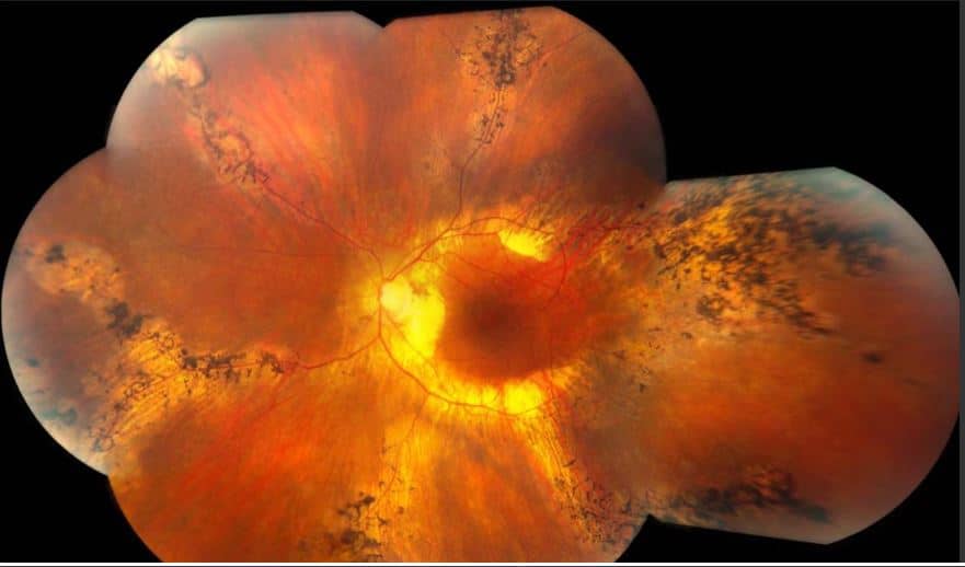
Ocular examination revealed visual acuity of 20/20 in the right eye and 20/40 in the left eye. Anterior segment examination and intraocular pressure were within normal limits in both eyes.
Smartphone fundoscopy of the left eye showed pigmented changes along retinal veins, extending to the choroid, with choroidal atrophy noted in the perivenous distribution.
Fluorescein angiography revealed hyperfluorescent staining along the affected veins without signs of leakage or neovascularization.
Optical coherence tomography demonstrated thinning of the choroid in perivenous areas and loss of the outer retinal layers.
A diagnosis of Pigmented Paravenous Retinochoroidal Atrophy (PPRCA) was made based on these characteristic findings.
DISEASE
Pigmented paravenous retinochoroidal atrophy (PPRCA) is a rare disorder of unknown etiology. The disease is characterized by pigment accumulation along the distribution of retinal veins. The findings are usually incidental with minimal effect on vision.
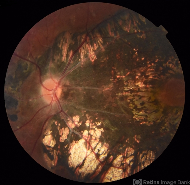
The diagnosis is made with a clinical examination due to the peculiar retinal findings.
The disease was first described in 1937 by Hewitson-Brown. The initial case was a 47-year-old male with past exposure to tuberculosis (multiple family members) who was admitted for alopecia areata.
An ophthalmic examination was remarkable for bilateral pigmentary changes along the distribution of the retinal vein.
Given the patient’s exposure to tuberculosis, Dr. Hewitson-Brown associated the retinal findings with tuberculosis. He termed the disease retino-choroiditis radiate.
Since then, it has appeared in the literature under multiple names including retinochoroiditis radiata, pseudoretinitis pigmentosa following measles, chorioeeretinitis striata, congenital pigmentation of the retina, melanosis of the retina, paravenous retinal degeneration, pigmented paravenous chorioretinal degeneration, pigmented paravenous chorioretinal atrophy and PPRCA.
The generally accepted name of the disease is either paravenous chorioretinal atrophy or pigmented paravenous retinochoroidal atrophy.
Fundus Explorer Pro
Photograph the retinal findings described here with the phone already in your pocket — 22 D optics and built-in illumination in one handheld unit.
From Choroida — the team behind this siteEtiology
The etiology of PPRCA remains unknown with a presumed initial inflammatory insult that leads to the slow development of the retinal findings.
Many inflammatory and infectious causes have been associated with the disease, including sarcoidosis, Behcet’s disease, syphilis, measles, rubeola, and tuberculosis.
However, no known systemic diseases have been identified as the cause of the retinal findings. The reported literature shows that the disease is more common in males than females.
There have been a few cases of familial occurrence, but currently, no known genes have been identified with the disease.

Diagnosis
Presentation:
The typical patient presents for an ophthalmologic exam and incidentally is found to have Pigmented paravenous retinochoroidal atrophy (PPRCA). The majority of patients are asymptomatic at presentation or have mild blurred vision.
The exception is patients with macular involvement, where vision can be severely reduced on presentation.
Physical examination:
PPRCA is an incidental finding and vision is rarely affected. Color vision is unaffected. Formal visual field testing may manifest scotomas along areas of densely involved retina. There are no specific findings on anterior and vitreous examination.
The funduscopic examination reveals bilateral involvement, with unilateral and asymmetric manifestations previously reported.
The retinal findings consist of bone corpuscle pigmentation, coarse pigment clumps, and fine pigmentary changes along the retinal veins.
The pigment accumulation and retinochoroidal atrophy begin a distance from the optic nerve head with the unaffected retina’s normal appearance.
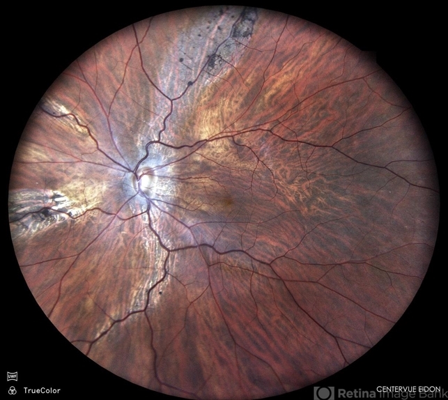
The pigment accumulation typically extends along the posterior pole and into the periphery. In marked cases, areas of retinal sheen are seen in the posterior pole.
Macular involvement is rare but cystoid macular edema, pigmentary macular degeneration, macular RPE atrophy, and lamellar macular holes have been reported.
The optic nerve appears normal with no gliosis or atrophy and the retinal vessel caliber is normal.
MANAGEMENT
PPRCA is a non-progressive or slowly progressive disease. There is no specific treatment for PPRCA.
Prognosis
Patients with PPRCA have a good visual prognosis with slowly progressive disease. In patients without macular involvement, visual acuity is minimally adversely affected.
Visual field loss does occur with time but at a rate slower than typical retinitis pigmentosa.


Document what you see
Two smartphone imaging tools built for everyday clinic use — one for the slit lamp, one for the fundus.
From Choroida — the team behind this siteREFERENCES
- Hewitson-Brown T. Retinochoroiditis radiata. Br J OphthalmoI1937;21:645. PMID: 18169492.
- Pigmented paravenous retinochoroidal atrophy (Review). Exp Ther Med. 2014 Jun; 7(6): 1439-1445. PMID: 24926324.
- Linek et al. Five cases of pigmented paravenous retinochoroidal atrophy in dogs. Veterinary Ophthalmology. 2012; 15. PMID: 22432693.
- Barteselli G. Fundus autofluorescence and optical coherence tomography findings in pigmented paravenous retinochoroidal atrophy. Can J Ophthalmology. 2014 Dec; 49(6):e144-6. PMID: 25433750.
- Ghosh B et al. SD-OCT in pigmented paravenous retinochoroidal atrophy. Ophthalmic Surg Lasers Imaging. 2012 May 10; 43(3):e41-3. PMID: 22589361.
- Batioglu F et al. Inflammatory pigmented paravenous retinochoroidal atrophy. Eye (London). 2002 Jan; 16(1):81-4. PMID: 11913895.
- Hashimoto Y et al. Abnormalities of fundus autofluorescence in pigmented paravenous chorioretinal atrophy. The Open Ophth J. 2012; 6: 125-128. PMID: 23264840.
- Fleckenstein M et al. Correlation of lines of increased autofluorescence in macular dystrophy and pigmented paravenous retinochoroidal atrophy by optical coherence tomography. Arch Ophthalmol. 2008 Oct; 126(10):1461-3. PMID: 18852430.
