
Salzmann Nodular Degeneration is one of those corneal findings that has usually been present and ignored for years.
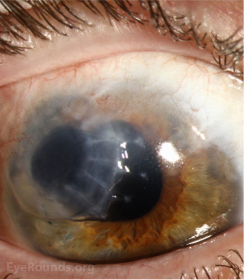
Elevated bluish-white nodules sit on the surface of an eye that has quietly endured chronic irritation.
They are easy to overlook until they blur vision or catch on every blink.
For clinicians, recognizing Salzmann Nodular Degeneration matters because it is a treatable, non-inflammatory degeneration that points back to an underlying ocular surface problem.
A careful slit-lamp look at the mid-peripheral cornea and a search for the surface disease behind it, is what the diagnosis needs.
What Is Salzmann Nodular Degeneration?
Salzmann Nodular Degeneration is a slowly progressive, non-inflammatory corneal degeneration characterised by elevated subepithelial nodules.
Its defining features are:
- Discrete, elevated, bluish-white to grey nodules
- A typically mid-peripheral or paracentral location, often in a ring
- Subepithelial fibrous and hyaline material between the epithelium and Bowman layer
- An association with chronic ocular surface disease
The nodules represent a reactive deposit rather than an active inflammation.
Epidemiology and Associations
It tends to appear on eyes with a long surface history.
- More common in older adults and in women
- Frequently linked to chronic surface disease — blepharitis, dry eye, and meibomian gland dysfunction
- Also associated with prior keratitis, trachoma, phlyctenular disease, and previous ocular surgery
The presence of a chronically irritated ocular surface is a recurring theme behind the nodules.
All-fit Slit-Lamp Adapter
Record and share exactly what you see at the slit lamp. One adapter fits any slit lamp or surgical microscope — and any smartphone.
From Choroida — the team behind this siteClinical Presentation
Symptoms
- Often asymptomatic and found incidentally
- Foreign-body sensation and irritation as the nodules elevate the epithelium
- Blurred vision and irregular astigmatism when the nodules approach the visual axis
- Recurrent epithelial breakdown over prominent nodules
Examination Findings
- Elevated bluish-white nodules, often several in a mid-peripheral arc
- A clear cornea between the nodules
- Signs of the underlying surface disease, such as blepharitis or dry eye
Discrete elevated bluish nodules on an otherwise clear cornea, in a chronically dry or inflamed eye, are the signature.
Diagnostic Evaluation
The diagnosis is clinical, supported by imaging when needed.
- Slit-lamp examination characterises the nodules and the surrounding cornea
- Anterior segment OCT shows the subepithelial deposit and measures its effect on the surface
- Corneal topography documents any induced irregular astigmatism
Anterior segment OCT is particularly useful for planning treatment when vision is affected.
Differential Diagnosis
A few surface lesions can resemble the nodules:
- Band keratopathy — calcific, in the interpalpebral cornea, not nodular
- Spheroidal (climatic droplet) degeneration — golden droplets rather than raised nodules
- Corneal scarring from prior keratitis — flat rather than elevated
- Ocular surface squamous neoplasia — gelatinous or leukoplakic, with a feeder vessel
The elevated, bluish, subepithelial nodules with a clear intervening cornea distinguish Salzmann degeneration from these mimics.
Management
Treatment starts with the surface and escalates only as needed.
- Lubrication and treatment of the underlying surface disease — dry eye and blepharitis — is first-line
- Superficial keratectomy, often a simple manual peel, removes visually significant or symptomatic nodules
- Phototherapeutic keratectomy can smooth the surface after nodule removal
- Nodules may recur, so ongoing surface care is important
Treating the ocular surface disease is what reduces symptoms and the risk of recurrence after removal.
Prognosis
The outlook is good.
- Vision usually recovers well after removal of central nodules
- The degeneration is benign and non-inflammatory
- Recurrence is possible, particularly if the surface disease is not controlled
In practice, the key to lasting improvement is treating the surface disease that drives the nodules, not just removing them.
Would you like to document corneal nodules and surface disease with your smartphone?


Document what you see
Two smartphone imaging tools built for everyday clinic use — one for the slit lamp, one for the fundus.
From Choroida — the team behind this siteReferences
- Werner LP, Issid K, Werner LP, et al. “Salzmann’s Nodular Corneal Degeneration.” Cornea. 2000;19(6):772–777.
- Maharana PK, Sharma N, Das S, et al. “Salzmann’s Nodular Degeneration.” The Ocular Surface. 2016;14(1):20–30.
- Graue-Hernandez EO, Mannis MJ, Eliasieh K, et al. “Salzmann Nodular Degeneration.” Cornea. 2010;29(3):283–289.
- American Academy of Ophthalmology. “Salzmann Nodular Degeneration.” EyeWiki.
Test yourself
A few questions straight from this article.
-
How is Salzmann nodular degeneration best characterised?
Salzmann nodular degeneration is a slowly progressive, non-inflammatory corneal degeneration in which the nodules represent a reactive deposit rather than active inflammation. -
Where does the fibrous and hyaline material of Salzmann nodular degeneration accumulate?
The nodules consist of subepithelial fibrous and hyaline material deposited between the corneal epithelium and Bowman layer. -
Where on the cornea do Salzmann nodules typically sit?
The nodules occupy a typically mid-peripheral or paracentral location, frequently arranged in a ring or arc, with clear cornea between them. -
Which chronic conditions are most frequently linked to Salzmann nodular degeneration?
Salzmann nodules are frequently linked to chronic ocular surface disease such as blepharitis, dry eye and meibomian gland dysfunction, alongside prior keratitis, trachoma and previous surgery. -
What role does anterior segment OCT play in Salzmann nodular degeneration?
Anterior segment OCT demonstrates the subepithelial deposit and its effect on the surface, and is particularly useful for planning treatment when vision is affected. -
Which feature distinguishes band keratopathy from Salzmann nodular degeneration?
Band keratopathy is a calcific change in the interpalpebral cornea and is not nodular, unlike the discrete elevated bluish nodules of Salzmann degeneration. -
Which appearance should raise suspicion of ocular surface squamous neoplasia rather than Salzmann nodules?
Ocular surface squamous neoplasia appears gelatinous or leukoplakic and carries a feeder vessel, unlike the elevated bluish subepithelial nodules of Salzmann degeneration. -
What is first-line management of Salzmann nodular degeneration?
Treatment starts with the ocular surface: lubrication and management of dry eye and blepharitis come first, with surgery reserved for nodules that remain symptomatic or visually significant. -
Which procedure removes visually significant or symptomatic Salzmann nodules?
Superficial keratectomy, often achieved as a simple manual peel, removes visually significant or symptomatic nodules, and phototherapeutic keratectomy can then smooth the surface. -
What most influences the risk of Salzmann nodules recurring after removal?
Vision usually recovers well after removal of central nodules, but recurrence is possible, particularly if the surface disease driving them is not controlled.
