AMD fellow eye describes the second eye of a patient who already has advanced macular degeneration in one eye.
This eye carries one of the highest conversion risks in macular practice, so every visit for the treated eye also serves as a screening visit for the other.
Why the AMD Fellow Eye Is High Risk
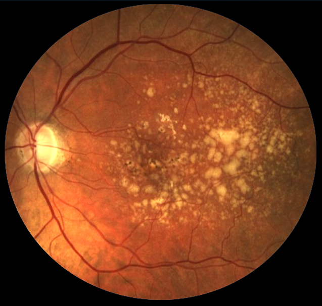
Both eyes share the same age, genes, smoking history and diet. As a result, drusen burden is usually symmetric, even when only one eye has converted.
In AREDS, participants with advanced AMD in one eye formed category 4, and this group showed the highest rates of progression during follow-up.
Phenotype of the First Eye
When the first eye has neovascular AMD, the fellow eye most often converts to neovascular disease. However, geographic atrophy can still develop, especially when reticular pseudodrusen are present. Therefore, record the phenotype of the first eye, because it shapes what you look for in the second.
Fundus Explorer Pro
Photograph the retinal findings described here with the phone already in your pocket — 22 D optics and built-in illumination in one handheld unit.
From Choroida — the team behind this siteGrading the AMD Fellow Eye at Baseline
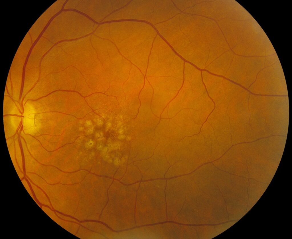
First, start with a dilated fundus exam and a color photograph, then grade drusen size and pigment changes (see AREDS severity scale). Large drusen and pigmentary abnormalities each add a risk point, and advanced AMD in the first eye already counts toward the person-level score.
OCT Findings That Raise Risk
Structural OCT shows changes that the fundus exam underestimates (see OCT biomarkers in AMD), and several features predict conversion in published cohorts:
- High drusen volume or large confluent drusen
- Also, intraretinal hyperreflective foci over drusen
- Reticular pseudodrusen (subretinal drusenoid deposits)
- In addition, hyporeflective cores within drusen
- Drusenoid pigment epithelial detachment
- Then, shallow irregular RPE elevation
Subclinical Neovascularization in the AMD Fellow Eye
OCT angiography can detect a vascular network under the RPE without any fluid, and swept-source OCTA studies described this nonexudative network in fellow eyes of unilateral exudative AMD.
As a result, these eyes showed a markedly higher risk of later exudation than eyes without a network. However, they did not need treatment while they stayed dry.
The Double-Layer Sign
A shallow irregular RPE elevation separated from Bruch membrane often marks a flat neovascular membrane, and OCTA can then confirm the flow signal. Meanwhile, injections are withheld unless fluid or hemorrhage appears.
Genetics and Lifestyle
CFH and ARMS2 risk alleles raise lifetime risk, but current guidance advises against routine genetic testing, because results do not change management today. Smoking also increases progression, so cessation advice belongs in every visit.
Monitoring Intervals for the AMD Fellow Eye
No single interval fits every patient: for example, many retina specialists image the fellow eye at each injection visit. However, when the treated eye moves to long extension intervals, the fellow eye may go months without OCT.
Therefore, set its own schedule, with shorter intervals for eyes carrying high-risk biomarkers, and book the next visit before a patient who stops injections leaves the clinic.
Symptoms the Patient Should Report
Ask about new distortion, a gray central patch or sudden blur. However, binocular viewing masks early change, because the brain favors the better image, so patients must test each eye separately.
Self-Checks for the AMD Fellow Eye
A daily monocular Amsler grid costs nothing and teaches patients what normal looks like, but its sensitivity is modest and adherence fades over time.
Preferential hyperacuity devices offer another option (see home monitoring for AMD), and in the HOME study, the device group had better acuity when neovascularization was detected.
Supplements for the AMD Fellow Eye
The AREDS formulation reduced progression to advanced AMD in high-risk participants, and then AREDS2 replaced beta carotene with lutein and zeaxanthin. This change matters for smokers, because beta carotene raises lung cancer risk.
Counseling About AMD Fellow Eye Conversion
Patients who lost vision in one eye often fear blindness, so first state the risk honestly and without exaggeration. Then explain that early treatment protects vision, because acuity at diagnosis strongly influences outcome.
Treating Conversion in the AMD Fellow Eye
Treat the new lesion with the same anti-VEGF approach used for any first eye. In addition, patients already in follow-up often present with smaller lesions and better acuity, and for newer agents, see new treatments for neovascular AMD.
Pitfalls to Avoid
- Do not skip fellow-eye OCT when the treated eye is stable
- Never rely on binocular symptom reports
- However, do not inject a dry eye with nonexudative neovascularization
- Also, avoid beta carotene in current or former smokers
- Do not lose patients who stop injections


Document what you see
Two smartphone imaging tools built for everyday clinic use — one for the slit lamp, one for the fundus.
From Choroida — the team behind this siteReferences
- Age-Related Eye Disease Study Research Group. A randomized trial of high-dose vitamins C and E, beta carotene, and zinc for age-related macular degeneration and vision loss: AREDS report no. 8. Arch Ophthalmol. 2001;119:1417-1436.
- Ferris FL, Davis MD, Clemons TE, et al. A simplified severity scale for age-related macular degeneration: AREDS report no. 18. Arch Ophthalmol. 2005;123:1570-1574.
- de Oliveira Dias JR, Zhang Q, Garcia JMB, et al. Natural history of subclinical neovascularization in nonexudative age-related macular degeneration using swept-source OCT angiography. Ophthalmology. 2018;125:255-266.
- Chew EY, Clemons TE, Bressler SB, et al. Randomized trial of a home monitoring system for early detection of choroidal neovascularization: the HOME study. Ophthalmology. 2014;121:535-544.
- Age-Related Eye Disease Study 2 Research Group. Lutein + zeaxanthin and omega-3 fatty acids for age-related macular degeneration: the AREDS2 randomized clinical trial. JAMA. 2013;309:2005-2015.

Writes and reviews the clinical articles on Educate. Every post is checked against peer-reviewed sources before it goes live.