Tear film layers describe the structure of the fluid that covers the ocular surface, from the outer lipid to the inner mucin.
Classic teaching names three layers: lipid, aqueous and mucin. However, current models treat the aqueous and mucin portions as one muco-aqueous gel, while each component fails differently and has its own bedside test.
What Are the Tear Film Layers?
The tear film coats the cornea and conjunctiva with a thin, stable fluid, and interferometry suggests a total thickness of only a few micrometers.
Because it smooths the corneal surface, it forms the first refractive interface of the eye, and it also lubricates the lids and carries antimicrobial proteins.
The Lipid Layer
Meibomian glands secrete the outermost lipid layer with each blink, with a thin polar phase facing the water and a thicker nonpolar phase facing air. Therefore, the layer slows evaporation and helps the film spread.
All-fit Slit-Lamp Adapter
Record and share exactly what you see at the slit lamp. One adapter fits any slit lamp or surgical microscope — and any smartphone.
From Choroida — the team behind this siteThe Aqueous Layer
The main and accessory lacrimal glands produce the aqueous component, which carries electrolytes, lysozyme, lactoferrin, secretory IgA and growth factors.
As a result, it nourishes the epithelium and defends against infection, while reflex tearing comes mainly from the main lacrimal gland.
The Mucin Layer
Corneal and conjunctival epithelia express membrane-bound mucins such as MUC1, MUC4 and MUC16, which form a glycocalyx that makes the cell surface wettable. In addition, conjunctival goblet cells secrete the gel-forming mucin MUC5AC, which traps debris.
Tear Film Layers and Dry Eye Classification
The TFOS DEWS II report places tear film instability and hyperosmolarity at the core of dry eye, and it describes evaporative and aqueous-deficient forms.
However, the two overlap, and many patients show features of both, so identifying the dominant failure guides treatment.
Lipid Failure: Evaporative Dry Eye
Meibomian gland dysfunction is the most common cause of evaporative dry eye, so at the slit lamp, look for capped orifices, lid margin telangiectasia and turbid meibum.
Patients often report burning that worsens through the day, and you should also consider reduced blinking during screen use and lagophthalmos.
Aqueous Failure in the Tear Film Layers
Aqueous deficiency follows reduced lacrimal secretion, and Sjögren syndrome is the classic cause, while age and systemic medications also contribute. Then the tear meniscus looks thin, and debris may float in it (see aqueous-deficient dry eye).
Mucin Failure on the Ocular Surface
Goblet cell loss leaves the epithelium poorly wettable, and cicatricial pemphigoid, Stevens-Johnson syndrome, chemical burns and vitamin A deficiency all reduce goblet cells. As a result, the film breaks up rapidly over areas that stain.
Testing the Lipid Layer
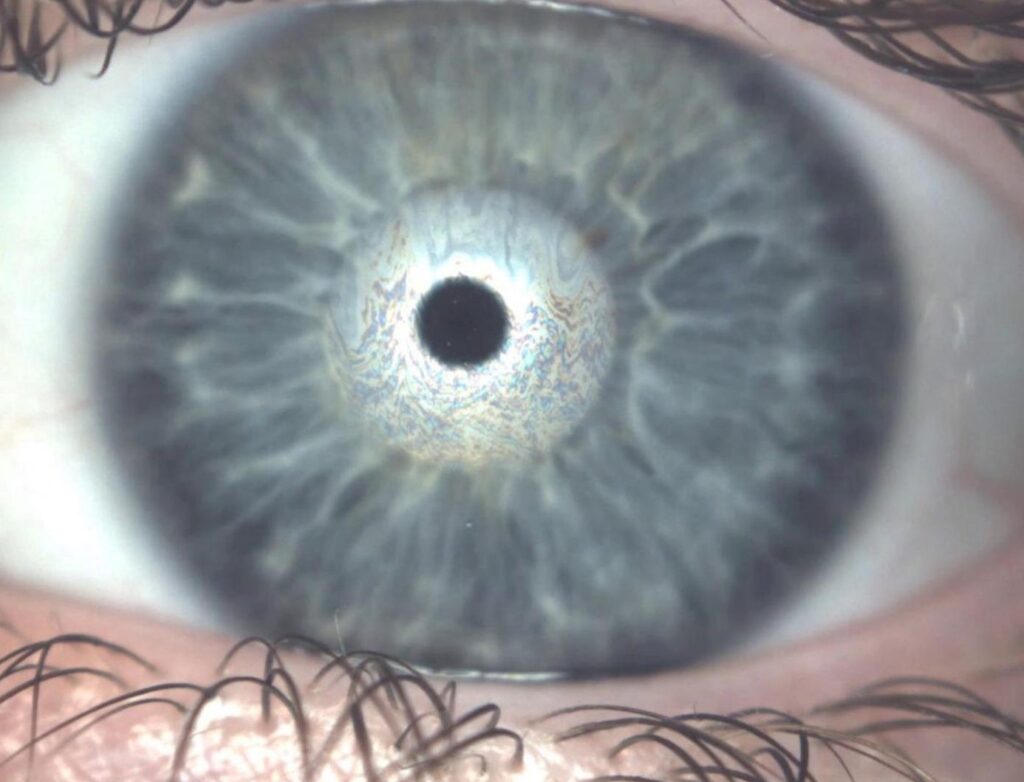
Gland expression shows meibum quality and gland function, while interferometry grades lipid layer thickness and meibography reveals gland dropout. In addition, noninvasive break-up time reflects lipid quality because evaporation drives early break-up.
Testing the Aqueous Layer
The Schirmer test measures strip wetting over five minutes (see Schirmer values), while tear meniscus height on slit lamp or anterior segment OCT offers a gentler estimate. However, both measures vary between visits.
Testing Mucin and Surface Wettability
Fluorescein break-up time shows how quickly the film destabilizes (see break-up time technique), and lissamine green stains cells that have lost their protective mucin coat.
Impression cytology counts goblet cells, although it is mainly a research tool, and tear osmolarity reflects global film failure, so it does not identify a single layer.
Reading the Pattern in Tear Film Layers
A short break-up time with a normal Schirmer result points toward evaporative disease. In contrast, a low Schirmer value with a thin meniscus suggests aqueous deficiency, and diffuse staining with rapid break-up despite normal volume raises concern for mucin damage.
Treatment Across the Tear Film Layers
For lipid failure, warm compresses, lid hygiene and in-office thermal treatment target the meibomian glands. For aqueous deficiency, options include preservative-free lubricants, topical cyclosporine or lifitegrast, and punctal plugs. Mucin damage requires control of the underlying disease first, and in addition, some countries approve diquafosol and rebamipide to stimulate mucin secretion.
Counseling Patients About Tear Film Layers
A simple analogy helps: oil on top, water in the middle and a sticky base, so patients then understand why compresses and drops address different problems. Finally, explain that treatment is long term because symptoms fluctuate.
Pitfalls to Avoid
- Do not treat every dry eye with drops alone; examine the lid margins first
- Measure tear meniscus and noninvasive break-up time before instilling fluorescein
- Record whether the Schirmer test used anesthetic
- Do not classify a patient on a single test
- Remember that reflex tearing can mask aqueous deficiency


Document what you see
Two smartphone imaging tools built for everyday clinic use — one for the slit lamp, one for the fundus.
From Choroida — the team behind this siteReferences
- Craig JP, Nichols KK, Akpek EK, et al. TFOS DEWS II definition and classification report. Ocul Surf. 2017;15:276-283.
- Willcox MDP, Argüeso P, Georgiev GA, et al. TFOS DEWS II tear film report. Ocul Surf. 2017;15:366-403.
- Bron AJ, de Paiva CS, Chauhan SK, et al. TFOS DEWS II pathophysiology report. Ocul Surf. 2017;15:438-510.
- Wolffsohn JS, Arita R, Chalmers R, et al. TFOS DEWS II diagnostic methodology report. Ocul Surf. 2017;15:539-574.
- Nichols KK, Foulks GN, Bron AJ, et al. The International Workshop on Meibomian Gland Dysfunction: executive summary. Invest Ophthalmol Vis Sci. 2011;52:1922-1929.
Test yourself
A few questions straight from this article.
-
In tear film layers of dry eye, which structures secrete the outermost lipid layer with each blink?
The article states that meibomian glands secrete the outermost lipid layer with each blink. The layer slows evaporation and helps the film spread. -
Which cells secrete the gel-forming mucin MUC5AC in the tear film that traps debris?
Conjunctival goblet cells secrete MUC5AC, a gel-forming mucin that traps debris. Membrane-bound mucins such as MUC1, MUC4 and MUC16 come from corneal and conjunctival epithelia. -
In current models of the tear film, which two components are treated as one muco-aqueous gel?
The article says current models combine the aqueous and mucin portions into one muco-aqueous gel, though each component fails differently and has its own bedside test. -
What is the most common cause of evaporative dry eye?
Meibomian gland dysfunction is the most common cause of evaporative dry eye. Look for capped orifices, lid margin telangiectasia and turbid meibum. -
Which cause is described as the classic cause of aqueous-deficient dry eye?
Aqueous deficiency follows reduced lacrimal secretion, and Sjögren syndrome is the classic cause. Age and systemic medications also contribute. -
Which finding in dry eye points toward evaporative disease rather than aqueous deficiency?
A short break-up time with a normal Schirmer result suggests evaporative disease. A low Schirmer value with a thin meniscus suggests aqueous deficiency. -
Which conditions reduce goblet cells and cause mucin failure in the tear film?
Cicatricial pemphigoid, Stevens-Johnson syndrome, chemical burns and vitamin A deficiency all reduce goblet cells, leaving the epithelium poorly wettable. -
In dry eye assessment, what does lissamine green staining highlight on the ocular surface?
Lissamine green stains cells that have lost their protective mucin coat. Meibography is what reveals gland dropout. -
Why does tear osmolarity not identify a single failing layer in dry eye?
The article states tear osmolarity reflects global film failure, so it does not identify a single layer. -
What treatment targets the meibomian glands in dry eye with lipid layer failure?
For lipid failure, warm compresses, lid hygiene and in-office thermal treatment target the meibomian glands. Punctal plugs, cyclosporine and lifitegrast are aqueous options.

Writes and reviews the clinical articles on Educate. Every post is checked against peer-reviewed sources before it goes live.