AMD vs DME is a frequent comparison, because both diseases blur central vision and thicken the macula.
Age-related macular degeneration (AMD) damages the outer retina, retinal pigment epithelium (RPE) and choroid. Diabetic macular edema (DME) is leakage from damaged retinal capillaries in diabetes, so history, fundus and OCT usually separate them within minutes.
Why Patients Confuse AMD vs DME
Both names contain the word “macular,” and both appear on the same injection schedule. Patients also hear “fluid” and “anti-VEGF” in both settings, and older diabetic patients can have both diseases at once.
The Patient Behind Each Disease
AMD typically affects people over 55, especially smokers and those with a family history. However, DME can occur at any age once diabetes has been present for several years, and poor glycemic control, hypertension and kidney disease raise its risk. Therefore, a young patient with type 1 diabetes and macular fluid is unlikely to have AMD.
Fundus Explorer Pro
Photograph the retinal findings described here with the phone already in your pocket — 22 D optics and built-in illumination in one handheld unit.
From Choroida — the team behind this siteSymptoms in AMD vs DME
Both groups report blur, distortion and difficulty reading. However, sudden distortion with a new central gray patch suggests neovascular AMD, while DME usually develops gradually, and early edema may cause no symptoms at all.
Fundus Signs of AMD
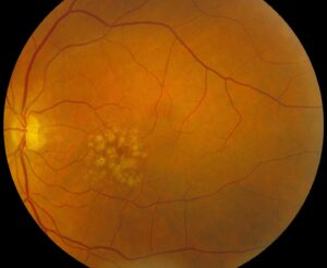
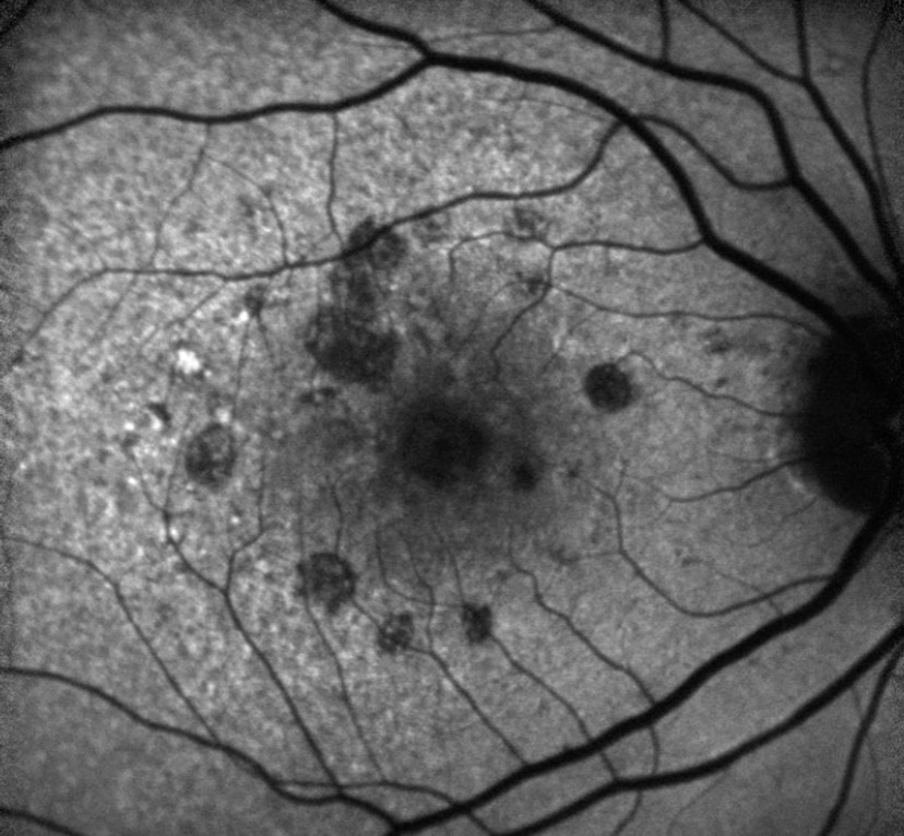
The hallmark is drusen, yellow deposits beneath the RPE, and pigment clumping, geographic atrophy and subretinal hemorrhage may also appear. Neovascular AMD can show a gray-green membrane, a lipid ring or a pigment epithelial detachment (PED), while the retinal vessels elsewhere typically look normal.
Fundus Signs of DME
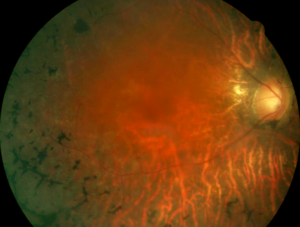
Here the examiner finds diabetic retinopathy around the macula, where microaneurysms, dot-blot hemorrhages and hard exudate rings are typical. Because DME complicates retinopathy, a wet macula without retinopathy elsewhere should prompt another diagnosis.
OCT Fluid Location in AMD vs DME
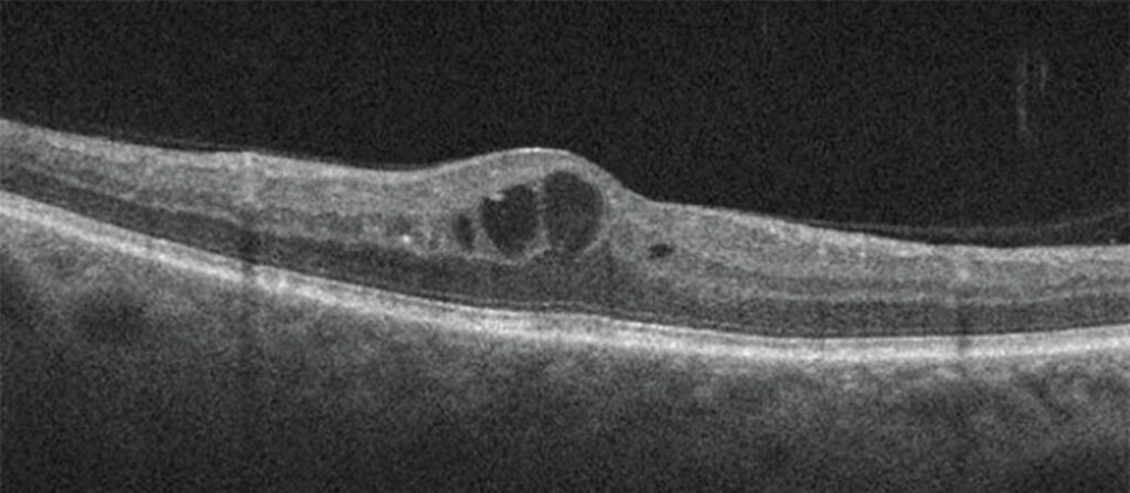
The layer holding the fluid is the most useful single clue. In neovascular AMD, fluid sits mainly under the retina or under the RPE, often above an irregular PED, although intraretinal cysts can also occur, especially with type 3 neovascularization.
In DME, by contrast, fluid fills cysts within the inner and outer nuclear layers (see DME OCT patterns). Hard exudates also appear as hyperreflective dots, while drusen form RPE bumps.
Angiography in AMD vs DME
Fluorescein angiography helps when OCT is ambiguous. Neovascular AMD leaks from a choroidal membrane, while DME leaks from microaneurysms and diffusely from capillaries. OCT angiography can also show a neovascular network in AMD and capillary dropout in DME.
Treatment Intent in AMD vs DME
The goals differ even when the drug is identical. In neovascular AMD, anti-VEGF therapy suppresses an active membrane that threatens rapid, permanent loss. In DME, anti-VEGF reduces leakage, while systemic control addresses the underlying vascular disease, so the DME plan always involves the diabetes team.
Anti-VEGF Evidence
MARINA showed that monthly ranibizumab prevented moderate vision loss in most eyes with neovascular AMD. Later, Protocol T compared aflibercept, bevacizumab and ranibizumab in center-involved DME, where all three improved vision, although aflibercept gained more when baseline vision was worse. However, injections control fluid and cannot restore photoreceptors lost to atrophy or fibrosis.
Other Options in DME
Steroid implants help eyes that respond poorly to anti-VEGF, particularly pseudophakic eyes, but they carry risks of cataract and raised intraocular pressure. Focal laser, first proven in the ETDRS, still treats selected leaks away from the center (see DME treatment choices).
Other Options in AMD
Dry AMD has no fluid to treat, but AREDS-based supplements lower the risk of progression in intermediate disease. Complement inhibitors now target geographic atrophy in some countries, and they slow lesion growth, yet they do not improve vision.
When AMD vs DME Overlap
Older diabetic patients can carry drusen and retinopathy together, so in that case the fluid source decides treatment. Subretinal fluid over a PED with a neovascular network favors AMD, whereas intraretinal cysts near microaneurysms favor DME.
Counseling Patients
Use plain words and first explain that AMD is wear in the support layer under the retina. DME, by contrast, is leakage from vessels damaged by diabetes, and stress that glucose and blood pressure control protect the diabetic eye. For AMD, add smoking cessation and home Amsler grid checks (see wet vs dry AMD).
Follow-Up
Both diseases need regular OCT review, and injection intervals should then follow fluid activity on OCT. Diabetic patients also need retinopathy screening even when vision feels normal.
Pitfalls to Avoid in AMD vs DME
- Labeling any macular fluid in a diabetic as DME without looking for drusen or a PED
- Missing polypoidal choroidal vasculopathy in an eye with hemorrhagic “wet AMD”
- Treating DME with injections alone while systemic control stays poor
- Promising visual recovery after fibrosis or atrophy


Document what you see
Two smartphone imaging tools built for everyday clinic use — one for the slit lamp, one for the fundus.
From Choroida — the team behind this siteReferences
- Rosenfeld PJ, Brown DM, Heier JS, et al. Ranibizumab for neovascular age-related macular degeneration. N Engl J Med. 2006;355:1419-1431.
- Wells JA, Glassman AR, Ayala AR, et al. Aflibercept, bevacizumab, or ranibizumab for diabetic macular edema. N Engl J Med. 2015;372:1193-1203.
- Early Treatment Diabetic Retinopathy Study Research Group. Photocoagulation for diabetic macular edema: ETDRS report number 1. Arch Ophthalmol. 1985;103:1796-1806.
- Age-Related Eye Disease Study 2 Research Group. Lutein + zeaxanthin and omega-3 fatty acids for age-related macular degeneration: the AREDS2 randomized clinical trial. JAMA. 2013;309:2005-2015.
- Spaide RF, Jaffe GJ, Sarraf D, et al. Consensus nomenclature for reporting neovascular age-related macular degeneration data. Ophthalmology. 2020;127:616-636.
Test yourself
A few questions straight from this article.
-
In diabetic macular edema, what is the underlying mechanism of macular thickening?
The article defines diabetic macular edema as leakage from damaged retinal capillaries in diabetes. AMD, by contrast, damages the outer retina, RPE and choroid. -
Which patient is most likely to have diabetic macular edema rather than age-related macular degeneration?
DME can occur at any age once diabetes has been present for several years, so a young patient with type 1 diabetes and macular fluid is unlikely to have AMD. -
Which symptom pattern suggests neovascular AMD rather than diabetic macular edema?
Sudden distortion with a new central gray patch suggests neovascular AMD, while DME usually develops gradually and early edema may cause no symptoms. -
Which funduscopic finding is the hallmark of age-related macular degeneration?
The article names drusen, yellow deposits beneath the RPE, as the AMD hallmark. Microaneurysms, dot-blot hemorrhages and hard exudate rings are typical of DME. -
A diabetic macular edema diagnosis is questionable when the macula is wet and what finding is absent?
Because DME complicates retinopathy, a wet macula without retinopathy elsewhere should prompt another diagnosis. -
On OCT, where does fluid mainly sit in neovascular AMD?
In neovascular AMD, fluid sits mainly under the retina or under the RPE, often above an irregular PED. Intraretinal cysts can occur, especially in type 3 neovascularization. -
On OCT, where is fluid typically found in diabetic macular edema?
In DME, fluid fills cysts within the inner and outer nuclear layers. Hard exudates appear as hyperreflective dots, while drusen form RPE bumps. -
How does fluorescein angiography leakage in diabetic macular edema differ from neovascular AMD?
Neovascular AMD leaks from a choroidal membrane, while DME leaks from microaneurysms and diffusely from capillaries. -
What did Protocol T show about anti-VEGF drugs in center-involved diabetic macular edema?
Protocol T compared aflibercept, bevacizumab and ranibizumab in center-involved DME. All three improved vision, but aflibercept gained more with worse baseline vision. -
What are the main risks of steroid implants used in diabetic macular edema?
Steroid implants help eyes responding poorly to anti-VEGF, particularly pseudophakic eyes, but they carry risks of cataract and raised intraocular pressure.

Writes and reviews the clinical articles on Educate. Every post is checked against peer-reviewed sources before it goes live.