PVD vs detachment is a daily triage question for anyone who examines patients with new floaters.
Both conditions can start with the same complaint, yet their urgency differs sharply, so the examiner needs a quick, reliable way to separate benign vitreous change from a threatening break.
Why PVD vs Detachment Is a Triage Question
Posterior vitreous detachment is a normal aging event in most adults, but vitreous traction during separation can tear the peripheral retina. A tear then lets fluid pass under the retina and produce a rhegmatogenous detachment.
As a result, every symptomatic PVD deserves a dilated peripheral examination.
How PVD Happens
The vitreous gel liquefies with age and collapses away from the retina. The posterior hyaloid usually separates first around the macula and then from the optic disc, while firm adhesion at the vitreous base stays intact for life.
Therefore, traction concentrates at the posterior border of the base, where most tears form. A flap tear then allows liquid vitreous to flow into the subretinal space. For background on the process, see our PVD overview.
Fundus Explorer Pro
Photograph the retinal findings described here with the phone already in your pocket — 22 D optics and built-in illumination in one handheld unit.
From Choroida — the team behind this siteSymptoms in PVD vs Detachment
Uncomplicated PVD typically causes a single large floater, cobwebs and brief temporal flashes, and vision stays clear apart from the moving opacity.
A retinal tear may add a sudden shower of many small dark dots. Detachment adds a fixed shadow, a curtain or distortion once the macula is involved.
Flashes and Floaters
Photopsia reflects mechanical traction on the retina, and patients notice it best in dim light. However, flashes alone do not separate a simple PVD from a tear.
A sudden increase in floaters, especially many dots, raises concern for bleeding from a torn vessel.
Visual Field Loss
Field loss marks the shift from a vitreous problem to a retinal problem. Patients describe a shadow that starts peripherally and creeps toward the center.
Inferior detachments often progress slowly, while superior ones can spread downward quickly. Because the macula may still be on, early referral protects central vision.
Shafer Sign in PVD vs Detachment
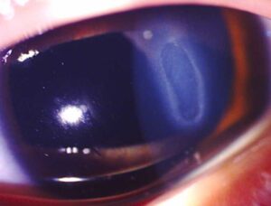
Shafer sign is pigment in the anterior vitreous, often called tobacco dust, and it represents retinal pigment epithelial cells released through a full thickness break. Tanner and colleagues found vitreous pigment a strong predictor of a break in acute PVD.
Vitreous hemorrhage carries similar weight, as Hollands and colleagues showed in their systematic review. Therefore, either finding demands a meticulous search for a tear (see horseshoe tears). If hemorrhage hides the periphery, B-scan ultrasound becomes essential at the first visit.
Examination Technique
First, record acuity and look for a relative afferent pupillary defect before dilation. Next, examine the anterior vitreous behind the lens with a narrow, bright slit beam.
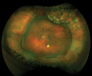
Then, use indirect ophthalmoscopy with scleral depression to view the vitreous base for 360 degrees. Finally, inspect any suspicious area with a three-mirror lens at the slit lamp.
Ultrasound and OCT in PVD vs Detachment
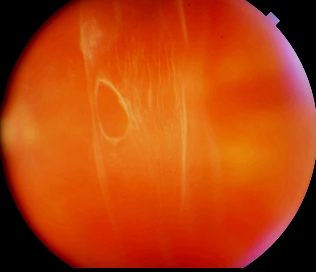
B-scan shows a thin, mobile posterior hyaloid in PVD and a thicker, tethered membrane in detachment. Also, a detached retina stays anchored to the optic disc, whereas a separated hyaloid may not.
OCT confirms partial separation over the macula and can reveal shallow subretinal fluid. However, OCT does not replace a peripheral examination.
Weiss Ring and Reassuring Signs
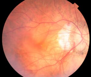
A Weiss ring over the disc confirms that the posterior hyaloid has separated from the nerve, and clear anterior vitreous, no hemorrhage and a flat periphery are also reassuring.
White without pressure is a common peripheral appearance and does not by itself signal a tear. Still, document these findings, because symptoms can evolve.
Same-Day Referral in PVD vs Detachment
The following findings justify same-day assessment by a retina specialist (see detachment triage):
- Shafer sign or any vitreous hemorrhage
- A visible retinal break or subretinal fluid
- A new curtain, shadow or field defect
- Reduced acuity or new distortion
- Dense hemorrhage that hides the periphery
- High myopia, lattice or a detachment in the fellow eye with new symptoms
Counseling After PVD vs Detachment Triage
Explain that floaters usually become less noticeable over months as the brain adapts. Then describe the warning signs clearly: a shower of new floaters, a shadow or reduced vision.
Ask the patient to return the same day if any of these appear. Written instructions also help, because dilation blurs vision during the discussion.
Follow-Up After Uncomplicated PVD
Some tears appear only after the initial examination, so the AAO Preferred Practice Pattern advises a repeat dilated examination within a few weeks.
Patients with hemorrhage or limited views may need closer review. Once the PVD is complete and the periphery stays flat, routine care resumes.
Pitfalls to Avoid
- Do not reassure a patient by phone without a dilated examination
- Never skip scleral depression in a symptomatic eye
- Do not ignore anterior vitreous pigment because the fundus looks normal
- Avoid attributing a field defect to floaters alone


Document what you see
Two smartphone imaging tools built for everyday clinic use — one for the slit lamp, one for the fundus.
From Choroida — the team behind this siteReferences
- Hollands H, Johnson D, Brox AC, et al. Acute-onset floaters and flashes: is this patient at risk for retinal detachment? JAMA. 2009;302:2243-2249.
- Tanner V, Harle D, Tan J, et al. Acute posterior vitreous detachment: the predictive value of vitreous pigment and symptomatology. Br J Ophthalmol. 2000;84:1264-1268.
- Flaxel CJ, Adelman RA, Bailey ST, et al. Posterior Vitreous Detachment, Retinal Breaks, and Lattice Degeneration Preferred Practice Pattern. Ophthalmology. 2020;127:P146-P181.
- Kang HK, Luff AJ. Management of retinal detachment: a guide for non-ophthalmologists. BMJ. 2008;336:1235-1240.
- Salmon JF. Kanski’s Clinical Ophthalmology: A Systematic Approach. 9th ed. Elsevier; 2020.

Writes and reviews the clinical articles on Educate. Every post is checked against peer-reviewed sources before it goes live.