High myopia is a refractive error of -6.00 D or worse, usually with an axial length of 26 mm or more.
As the eye elongates, the sclera, choroid and retina thin and stretch, and these changes raise the lifetime risk of several sight-threatening diseases. Holden and colleagues projected nearly one billion people with high myopia by 2050.
How High Myopia Is Defined
The International Myopia Institute defines high myopia as a spherical equivalent of -6.00 D or worse with accommodation relaxed. However, some epidemiological reports, including WHO work, use -5.00 D, and many clinicians add an axial length threshold of 26 mm. Therefore, always note which definition a study or chart uses.
High Myopia and Pathologic Myopia
High myopia describes the degree of refractive error, while pathologic myopia describes structural damage to the posterior segment. The META-PM system defines it as diffuse atrophy or worse, plus lesions, or posterior staphyloma. However, many highly myopic eyes stay healthy for decades.
Fundus Explorer Pro
Photograph the retinal findings described here with the phone already in your pocket — 22 D optics and built-in illumination in one handheld unit.
From Choroida — the team behind this siteRetinal Detachment Risk in High Myopia
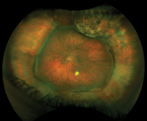
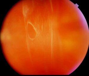
Long eyes develop lattice degeneration, atrophic holes and earlier posterior vitreous detachment, and vitreous traction then tears the thin peripheral retina more easily. Pooled data show detachment risk rising steeply with the degree of myopia.
Repair is also harder in these eyes (see retinal detachment in high myopia), and macular holes in staphylomatous eyes can cause posterior pole detachment (see macular hole retinal detachment).
Glaucoma in High Myopia
Myopia is an established risk factor for open-angle glaucoma. A meta-analysis by Marcus and colleagues confirmed the association, with higher odds in more myopic eyes. Because the lamina cribrosa is thin and stretched, it may tolerate pressure poorly.
Why Glaucoma Diagnosis Is Difficult
Tilted discs and peripapillary atrophy obscure the true neuroretinal rim, and OCT normative databases often exclude long eyes, so false red flags are common. Staphyloma and atrophy can also produce field defects without glaucoma, so base the diagnosis on documented progression over time.
Cataract in High Myopia
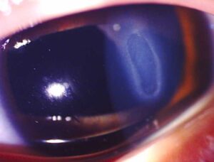
Highly myopic eyes develop nuclear and posterior subcapsular cataract at a younger age, and patients often notice a myopic shift before glare becomes obvious. Biometry needs care, because staphyloma can distort axial length readings. Surgery also raises the later retinal detachment risk, especially in younger men.
Myopic Maculopathy in High Myopia
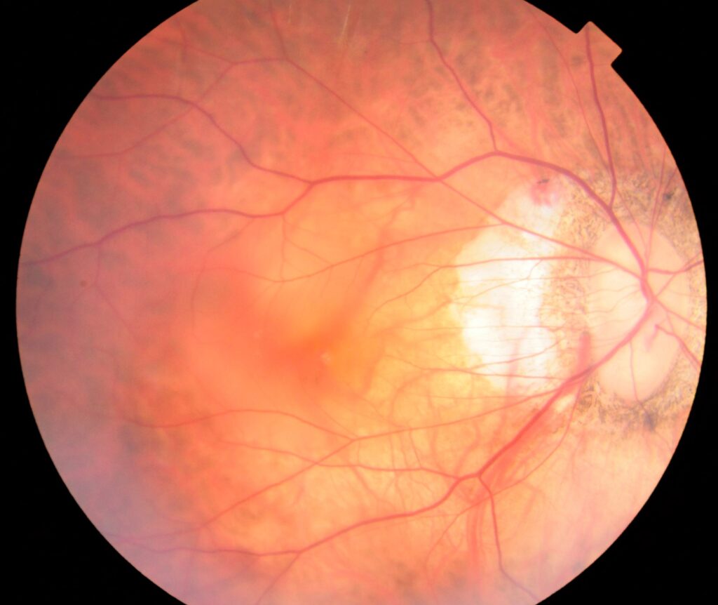
Maculopathy is a leading cause of irreversible vision loss in this group. Changes progress from tessellation to diffuse and patchy atrophy, then macular atrophy. Lacquer cracks and Fuchs spots are plus lesions that signal higher risk.
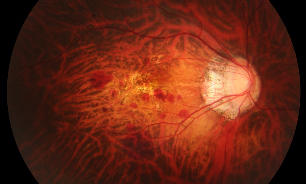
Myopic Choroidal Neovascularization
Myopic CNV is usually small, subfoveal and type 2, and patients report sudden distortion or a small central scotoma. Anti-VEGF therapy is first-line, and RADIANCE showed better gains with ranibizumab than verteporfin therapy. However, atrophy can expand around the scar years later.
Myopic Traction Maculopathy
OCT reveals foveoschisis, lamellar holes or foveal detachment in staphylomatous eyes, and many cases stay stable for long periods. However, progression to a full-thickness hole or foveal detachment may justify vitrectomy, so serial OCT guides timing better than one image.
Exam Checklist for High Myopia
- Refraction, with cycloplegia in children and young adults
- Axial length by optical biometry
- Intraocular pressure and central corneal thickness
- Slit-lamp lens grading and history of myopic shift
- Disc photos, RNFL OCT and baseline visual fields
- Macular OCT for schisis, CNV and holes
- Dilated peripheral exam with scleral depression
Follow-Up in High Myopia
Most adults need a dilated examination at least yearly. However, maculopathy, glaucoma suspicion or recent cataract surgery shortens the interval. Next, compare OCT and field results with the patient’s own baseline.
Counseling Patients With High Myopia
Explain that glasses, contacts and refractive surgery correct focus only, and none of them shortens the eye or removes the retinal risk. Then teach the warning signs: new floaters, flashes or a curtain (see retinal detachment symptoms), and also advise monocular checks for new distortion.
Preventing High Myopia in Children
Children who become myopic early are more likely to reach high myopia, so myopia control has the most value in young patients. Options include low-dose atropine, orthokeratology, defocus spectacles and more outdoor time, and these tools slow elongation, while existing axial length does not reverse.
Pitfalls to Avoid
- Do not call a large cup glaucoma before repeating OCT and fields
- Never skip scleral depression in symptomatic highly myopic patients
- Avoid assuming that LASIK removed retinal risk
- Exclude CNV before blaming new central loss on atrophy


Document what you see
Two smartphone imaging tools built for everyday clinic use — one for the slit lamp, one for the fundus.
From Choroida — the team behind this siteReferences
- Flitcroft DI, He M, Jonas JB, et al. IMI, defining and classifying myopia: a proposed set of standards for clinical and epidemiologic studies. Invest Ophthalmol Vis Sci. 2019;60:M20-M30.
- Holden BA, Fricke TR, Wilson DA, et al. Global prevalence of myopia and high myopia and temporal trends from 2000 through 2050. Ophthalmology. 2016;123:1036-1042.
- Ohno-Matsui K, Kawasaki R, Jonas JB, et al. International photographic classification and grading system for myopic maculopathy. Am J Ophthalmol. 2015;159:877-883.
- Marcus MW, de Vries MM, Junoy Montolio FG, Jansonius NM. Myopia as a risk factor for open-angle glaucoma: a systematic review and meta-analysis. Ophthalmology. 2011;118:1989-1994.
- Flitcroft DI. The complex interactions of retinal, optical and environmental factors in myopia aetiology. Prog Retin Eye Res. 2012;31:622-660.
- Wolf S, Balciuniene VJ, Laganovska G, et al. RADIANCE: a randomized controlled study of ranibizumab in patients with choroidal neovascularization secondary to pathologic myopia. Ophthalmology. 2014;121:682-692.

Writes and reviews the clinical articles on Educate. Every post is checked against peer-reviewed sources before it goes live.