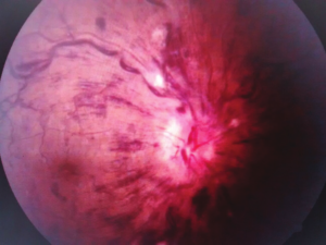
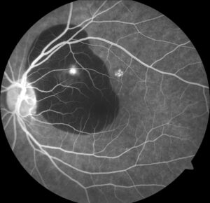
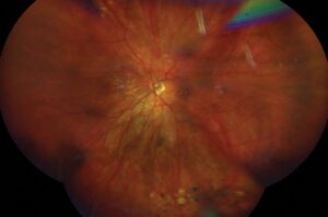
Retinal detachment symptoms are a combination of flashes of light, new floaters, and a shadow or curtain across part of the vision.
A patient who describes them needs to be seen quickly, because early treatment of a tear or a detachment gives the best chance of keeping vision.
Not every patient with floaters has a detachment, and good triage separates the urgent from the routine.
Retinal Detachment Symptoms Patients Describe
- Photopsia: brief flashes of light, usually in the peripheral field, seen best in the dark, caused by traction of the vitreous on the retina
- Floaters: new dots, threads, cobwebs, or a ring, which may also appear suddenly
- A shower of floaters: many new dark spots, which suggests vitreous hemorrhage or pigment cells from a tear
- A shadow or curtain: a dark area in the peripheral field that spreads, a sign of detachment
- Reduced central vision or distortion, which suggests macular involvement
Symptoms are usually in one eye, and the patient may notice them especially against a bright background.
Retinal Detachment Symptoms: Why Timing Matters
A posterior vitreous detachment occurs in most people with age, but it is usually harmless (see posterior vitreous detachment).
In a minority, the vitreous pulls a retinal tear as it separates, and the tear can also lead to a detachment over days or weeks.
Treating a tear with laser or cryotherapy is simple and effective, whereas repairing a detachment requires surgery, and detachment involving the macula has a worse visual prognosis (see macula-off retinal detachment).
Fundus Explorer Pro
Photograph the retinal findings described here with the phone already in your pocket — 22 D optics and built-in illumination in one handheld unit.
From Choroida — the team behind this siteFeatures That Raise Urgency
- Sudden onset of floaters with flashes
- A shower of new floaters, or a sudden increase in the number
- A visible field defect or shadow that is enlarging
- Reduced vision or distortion of recent onset
- Recent eye surgery or trauma
- High myopia, a history of detachment in the other eye, or a family history
- Previous cataract surgery or lattice degeneration
Patients with these features need examination on the same day, or within 24 hours at the most.
Retinal Detachment Symptoms: Telephone and Primary Care Triage
- Ask when the symptoms started, whether they are new, and whether they are getting worse
- First, ask about flashes, a shower of floaters, a shadow, and changes in central vision
- Question about risk factors, such as myopia, surgery, and previous detachment
- Give clear advice: new flashes and floaters, or a shadow, need an examination within a day
- Also advise patients with a curtain or sudden loss of vision to go immediately to an eye service
Written protocols help nurses and optometrists to decide, while a clear route for urgent referral prevents delays.
What the Examination Includes
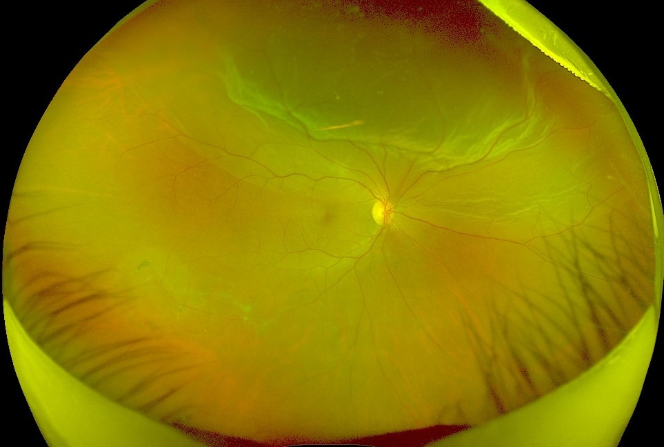
- Visual acuity, pupils, and visual field by confrontation
- A dilated examination of the retina, with scleral depression (see scleral depression)
- Examination of the vitreous for pigment cells, hemorrhage, and Weiss ring
- OCT of the macula when there are visual symptoms
- B-scan ultrasonography if the view is poor (see B-scan ultrasonography)
- Examination of the other eye
A retinal tear is treated the same day when possible, and patients with a detachment are scheduled for surgery.
The Risk of Delayed Tears
Even when the first examination is normal, a small proportion of patients with a symptomatic posterior vitreous detachment develop a new tear in the following weeks.
Coffee and colleagues found that about 3 to 4 percent of patients with a negative initial examination developed delayed breaks (see the references).
Van Overdam and colleagues found that the presence of pigment cells in the vitreous, vitreous hemorrhage, and certain symptoms predicted the development of new breaks (see the references).
For this reason, follow-up at about four to six weeks is usually advised, and patients are told to return at once if symptoms change.
Counselling After a Normal Examination
- Explain that the symptoms are due to the vitreous gel pulling away, that no tear was found, and that it is a common benign process
- Then teach the warning signs: more floaters, more flashes, a shadow, or reduced vision
- Arrange a follow-up visit in four to six weeks, since delayed breaks can appear
- Give a telephone number and tell the patient to call at any time if these changes occur
A clear plan also reassures patients and protects them.
A Simple Triage Table
- Same day: flashes with new floaters, a shadow or curtain, a sudden drop in vision, a recent injury or surgery
- Within one to two days: new floaters without flashes, in a patient with risk factors
- Routine review: longstanding floaters that have not changed
Use this as a guide, and when in doubt, choose the more urgent option.
For the latest studies, see the PubMed results for retinal detachment symptoms.


Document what you see
Two smartphone imaging tools built for everyday clinic use — one for the slit lamp, one for the fundus.
From Choroida — the team behind this siteReferences
- Coffee RE, Westfall AC, Davis GH, Mieler WF, Holz ER. Symptomatic posterior vitreous detachment and the incidence of delayed retinal breaks: case series and meta-analysis. Am J Ophthalmol. 2007;144:409-413.
- van Overdam KA, Bettink-Remeijer MW, Mulder PG, van Meurs JC. Symptoms and findings predictive for the development of new retinal breaks. Arch Ophthalmol. 2005;123:479-484.
- Ryan SJ, Schachat AP, Wilkinson CP, et al, eds. Retina. 6th ed. Elsevier; 2018.
- American Academy of Ophthalmology. Basic and Clinical Science Course, Section 12: Retina and Vitreous. AAO; current edition.

Writes and reviews the clinical articles on Educate. Every post is checked against peer-reviewed sources before it goes live.