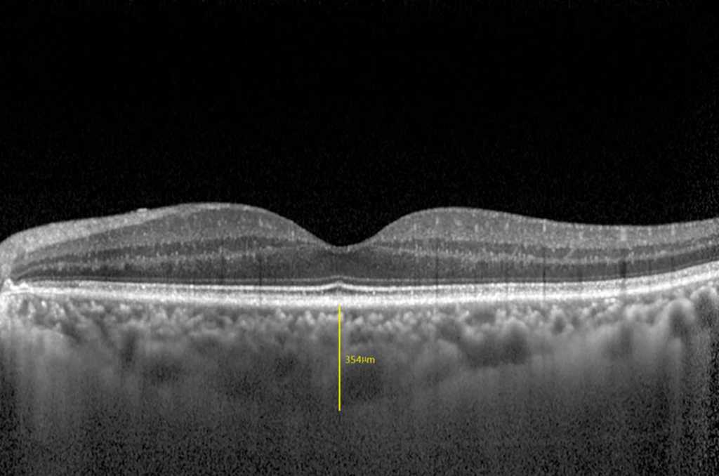
Choroidal thickness is the distance from the outer border of the retinal pigment epithelium to the chorioscleral interface.
Enhanced depth imaging OCT made this measurement routine.
Therefore, clinicians can now follow the choroid in many diseases.
Why Measure Choroidal Thickness
The choroid supplies the outer retina and shapes many diseases.
Thickening and thinning mark different disease groups.
Also, treatment can change the thickness.
As a result, the measure adds to the diagnosis and follow-up.
Enhanced Depth Imaging
Spaide and colleagues described enhanced depth imaging in 2008.
The device is placed close to the eye so that the choroid sits near the zero delay line.
Also, the technique increases sensitivity in the deeper layers.
Therefore, standard spectral-domain machines can show the choroid.
Fundus Explorer Pro
Photograph the retinal findings described here with the phone already in your pocket — 22 D optics and built-in illumination in one handheld unit.
From Choroida — the team behind this siteSwept-Source OCT
Swept-source devices use a longer wavelength that penetrates deeper.
They image the choroid and the sclera without special settings.
Also, the signal is stronger through cataract and blood.
As a result, many centers now prefer swept-source scans.
Measuring Subfoveal Choroidal Thickness
Place a caliper at the outer border of the pigment epithelium and drag it to the inner scleral border.
Take the measurement under the center of the fovea.
Also, repeat at points 500 and 1,000 micrometers nasal and temporal.
Therefore, report the subfoveal value and the location.
Normal Choroidal Thickness Values
Margolis and Spaide found a mean subfoveal thickness of about 287 micrometers in normal eyes.
Values fall with age and with axial length.
Also, the range is wide among healthy people.
As a result, use age- and refraction-matched reference data.
Effect of Age and Myopia on Choroidal Thickness
The choroid thins by about 1.5 micrometers each year of age.
It also thins with each diopter of myopia.
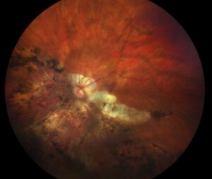
Also, high myopia can reduce thickness to under 100 micrometers (see tessellated fundus).
Therefore, interpret thickness in context.
Diurnal and Other Variations
Brown and colleagues showed that thickness changes through the day.
The choroid is thickest in the morning and thinner in the evening.
Also, caffeine, water intake and exercise can alter it.
In addition, schedule repeat scans at the same time of day.
Pachychoroid Disease
Thick choroid with dilated large vessels defines the pachychoroid spectrum.
Central serous chorioretinopathy, pachychoroid neovasculopathy and polypoidal vasculopathy are in the group.
Also, thickness often exceeds 350 micrometers in affected eyes.
Therefore, a thick choroid should prompt a search for these diseases.
Thin Choroid in AMD and Myopia
Eyes with geographic atrophy and pathologic myopia show thin choroid.
The thinning may precede visible atrophy.
Also, thin choroid predicts poor response in some diseases.
As a result, thinness serves as a marker of risk.
Inflammatory Disease
Choroiditis such as Vogt-Koyanagi-Harada disease thickens the choroid in the acute phase.
Treatment with steroids reduces the thickness.
Also, serial scans help to track recovery.
In addition, thickness can warn of relapse.
Choroidal Vascularity Index
Agrawal and colleagues proposed the ratio of vessel area to total choroid area.
It separates vessels from stroma in binarized images.
Also, it is less sensitive to age and refraction than thickness.
Therefore, it adds a second measure of choroidal status.
Choroidal Thickness After Treatment
Photodynamic therapy thins the choroid in central serous disease.
Anti-VEGF therapy can reduce thickness in some eyes.
Also, mineralocorticoid antagonists may thin it in selected patients.
As a result, thickness tracks a response.
Pitfalls in Measuring Choroidal Thickness
The outer border can be hard to see in thick or thin choroid.
Shadowing from pigment or hemorrhage blurs the line.
Also, manual caliper errors affect small changes.
Therefore, use the same observer and the same method.
Automated Segmentation
Software can segment the choroid and give maps.
Errors occur in eyes with disease, so review the lines.
Also, maps allow comparison across the macula.
In addition, report the device and software version.
Clinical Use Today
Use thickness to support a diagnosis and to track disease.
Do not treat on a thickness value alone.
Also, combine it with fluid, angiography and symptoms.
As a result, the measure serves as an adjunct.
Pitfalls to Avoid
- Do not compare values between devices without correction
- Never interpret thickness without age and refraction
- Do not forget the time of day in serial scans
- Never use thickness alone to start treatment
Reporting Choroidal Measurements
State the device, the scan mode and the point of measurement.
Give the date and time of the scan.
Also, include age and refraction.
Therefore, others can compare your numbers with yours.
For the latest studies, see the PubMed results for choroidal thickness.


Document what you see
Two smartphone imaging tools built for everyday clinic use — one for the slit lamp, one for the fundus.
From Choroida — the team behind this siteReferences
- Spaide RF, Koizumi H, Pozzoni MC. Enhanced depth imaging spectral-domain optical coherence tomography. Am J Ophthalmol. 2008;146:496-500.
- Margolis R, Spaide RF. A pilot study of enhanced depth imaging optical coherence tomography of the choroid in normal eyes. Am J Ophthalmol. 2009;147:811-815.
- Brown JS, Flitcroft DI, Ying GS, et al. In vivo human choroidal thickness measurements: evidence for diurnal fluctuations. Invest Ophthalmol Vis Sci. 2009;50:5-12.
- Agrawal R, Gupta P, Tan KA, Cheung CM, Wong TY, Cheng CY. Choroidal vascularity index as a measure of vascular status of the choroid: measurements in healthy eyes from a population-based study. Sci Rep. 2016;6:21090.
Test yourself
A few questions straight from this article.
-
How is choroidal thickness defined on OCT?
Choroidal thickness is the distance from the outer border of the retinal pigment epithelium to the chorioscleral interface. -
How does enhanced depth imaging OCT bring the choroid into view?
With EDI the device is placed close to the eye so the choroid sits near the zero delay line, raising sensitivity in deeper layers on standard spectral-domain machines. -
How should a choroidal thickness value be used in treatment decisions?
Thickness supports diagnosis and tracks disease, but treatment should never be started on a thickness value alone; combine it with fluid, angiography and symptoms. -
What mean subfoveal choroidal thickness did Margolis and Spaide find in normal eyes?
Margolis and Spaide found a mean subfoveal thickness of about 287 micrometers in normal eyes, with a wide healthy range. -
By roughly how much does the choroid thin with each year of age?
The choroid thins by about 1.5 micrometers each year of age and also thins with each diopter of myopia, so age- and refraction-matched data are needed. -
Subfoveal choroidal thickness in pachychoroid disease often exceeds which value?
Thick choroid with dilated large vessels defines the pachychoroid spectrum, and thickness often exceeds 350 micrometers in affected eyes. -
Which conditions are associated with a thin choroid on EDI-OCT?
Eyes with geographic atrophy and pathologic myopia show a thin choroid, and the thinning may precede visible atrophy. -
How does acute Vogt-Koyanagi-Harada disease affect choroidal thickness?
Choroiditis such as VKH thickens the choroid in the acute phase, steroid treatment reduces the thickness, and serial scans track recovery and warn of relapse. -
What does the choroidal vascularity index measure?
The choroidal vascularity index is the ratio of vessel area to total choroid area on binarized images, and it is less sensitive to age and refraction than thickness. -
Why should serial choroidal thickness scans be taken at the same time of day?
Brown and colleagues showed diurnal change in choroidal thickness, and caffeine, water intake and exercise can alter it, so repeat scans at the same time of day.

Writes and reviews the clinical articles on Educate. Every post is checked against peer-reviewed sources before it goes live.