PVD stages describe how the posterior vitreous separates from the retina over months to years.
Optical coherence tomography (OCT) shows this process long before most patients notice floaters.
Therefore, staging helps clinicians link a scan to symptoms, risk and follow-up.
Why PVD Stages Exist
Clinicians once described PVD simply as present or absent.
However, OCT showed that separation usually starts near the fovea and spreads slowly.
Uchino and colleagues described these early steps in healthy older eyes.
Later, Johnson linked each step to specific vitreomacular disease.
Vitreous Anatomy Before Separation
The posterior hyaloid adheres most firmly at the vitreous base, optic disc, fovea and retinal vessels.
With age, the gel liquefies (see vitreous syneresis).
At the same time, adhesion at the vitreoretinal interface weakens.
As a result, liquid vitreous gains access to the potential space between cortex and retina.
Fundus Explorer Pro
Photograph the retinal findings described here with the phone already in your pocket — 22 D optics and built-in illumination in one handheld unit.
From Choroida — the team behind this siteStage 0: Attached Vitreous
In stage 0, the posterior hyaloid lies flat on the retina.
OCT shows no separate hyaloid line over the macula.
However, a flat retina can also follow complete separation, so context matters.
Stage 1 in PVD Stages: Focal Perifoveal Separation
Stage 1 begins with a shallow perifoveal detachment in one or more quadrants, but not all.
The hyaloid lifts as a thin line, then dips back to the retina.
Patients rarely notice anything at this point.
Also, the stage can persist unchanged for a long time.
Stage 2: Separation Around the Fovea
In stage 2, the perifoveal detachment involves every quadrant.
The hyaloid remains attached at the foveal center and the optic disc.
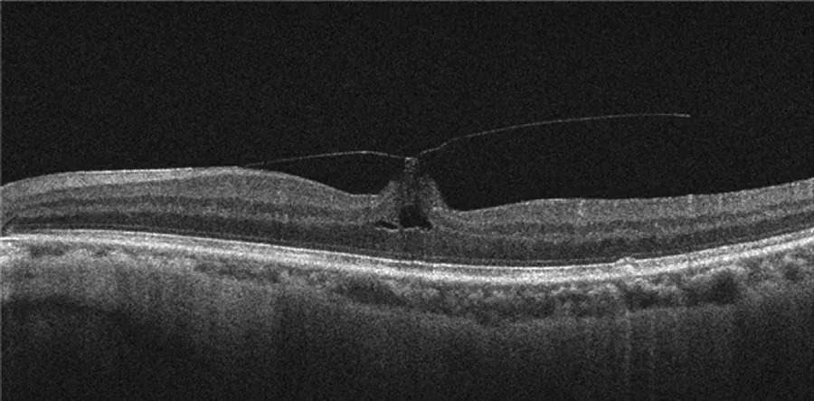
On OCT, it forms a gull-wing profile over the macula.
Therefore, traction concentrates on a small foveal point.
Stage 3: Release From the Fovea
Stage 3 starts when the hyaloid lets go of the fovea.
However, it still clings to the optic disc.
OCT shows a hyaloid line floating freely above the macula.
In contrast, a wide scan or ultrasound may reveal attachment at the disc margin.
Stage 4: Complete PVD and the Weiss Ring
Stage 4 marks complete separation, including release from the optic disc.
The detached peripapillary ring often appears as a floating Weiss ring at the slit lamp (see Weiss ring).
Then, OCT may show no hyaloid within the scan window.
Many patients notice floaters or flashes during this final step.
Comparing PVD Stages Side by Side
- Attached (stage 0): no hyaloid line, no symptoms
- Focal perifoveal (stage 1): partial separation, fovea and disc attached, usually silent
- Full perifoveal (stage 2): fovea and disc attached, possible foveal traction
- Foveal release (stage 3): disc still attached, usually silent
- Complete (stage 4): Weiss ring possible, floaters and flashes common
Reading PVD Stages on OCT
First, use long horizontal and vertical line scans through the fovea.
Next, widen the scan to include the optic disc.
Also, raise contrast so the faint hyaloid stands out.
Finally, compare both eyes, because separation is often bilateral but asymmetric.
Limits of OCT for PVD Stages
Standard OCT images only the posterior pole.
As a result, it can miss peripheral or peripapillary separation.
In addition, the posterior wall of the premacular bursa can mimic a detached hyaloid.
Dynamic B-scan ultrasound often clarifies whether the hyaloid moves freely.
Symptoms Across PVD Stages
Early stages are usually painless and visually quiet.
Symptoms tend to cluster around stage 4, when the gel collapses.
Patients describe a new floater, cobwebs or brief temporal flashes.
However, some complete separations pass without any noticeable event.
When Separation Turns Pathologic
Anomalous PVD occurs when the gel liquefies faster than adhesion weakens.
For example, persistent foveal traction in stage 2 can produce vitreomacular traction.
Then, a full-thickness hole may follow (see macular hole staging).
Peripheral traction during stage 4 can tear the retina and cause hemorrhage.
Follow-up Decisions by PVD Stages
Asymptomatic stage 1 to 3 findings need no treatment.
Acute symptomatic stage 4 needs a dilated peripheral examination with scleral depression.
Hollands and colleagues found that vitreous hemorrhage or pigment raised the likelihood of a tear.
Therefore, many clinicians repeat the peripheral examination within weeks when findings are uncertain.
Counseling Patients About PVD Stages
Explain that separation is a normal aging change in most eyes.
Also, mention that early stages may progress silently.
Teach the warning signs: a shower of floaters, a curtain or persistent flashes.
Then, advise urgent review if they appear.
Pitfalls to Avoid
- Do not call a flat hyaloid stage 4 without ultrasound or a Weiss ring
- Never judge disc attachment from a narrow macular scan
- Do not dismiss stage 2 traction when the foveal contour changes
- Avoid reassurance before a full peripheral examination in acute symptoms


Document what you see
Two smartphone imaging tools built for everyday clinic use — one for the slit lamp, one for the fundus.
From Choroida — the team behind this siteReferences
- Uchino E, Uemura A, Ohba N. Initial stages of posterior vitreous detachment in healthy eyes of older persons evaluated by optical coherence tomography. Arch Ophthalmol. 2001;119:1475-1479.
- Johnson MW. Posterior vitreous detachment: evolution and complications of its early stages. Am J Ophthalmol. 2010;149:371-382.
- Duker JS, Kaiser PK, Binder S, et al. The International Vitreomacular Traction Study Group classification of vitreomacular adhesion, traction, and macular hole. Ophthalmology. 2013;120:2611-2619.
- Hollands H, Johnson D, Brox AC, et al. Acute-onset floaters and flashes: is this patient at risk for retinal detachment? JAMA. 2009;302:2243-2249.
- Sebag J. Anomalous posterior vitreous detachment: a unifying concept in vitreo-retinal disease. Graefes Arch Clin Exp Ophthalmol. 2004;242:690-698.

Writes and reviews the clinical articles on Educate. Every post is checked against peer-reviewed sources before it goes live.