Vitreous syneresis is the process in which the vitreous gel loses its structure, the collagen fibrils clump together, and pockets of liquid form between them.
It is a normal part of aging, and it is the usual starting point for floaters, for posterior vitreous detachment, and for several of the traction problems that follow.
Patients rarely come to the clinic because of the liquefaction itself, but because of what they see as a result.
What Is Vitreous Syneresis?
The healthy vitreous is a transparent gel made mainly of type II collagen fibrils, hyaluronan, and water.
The hyaluronan keeps the collagen fibrils separated, and this separation is what keeps the gel clear.
With age, hyaluronan molecules and collagen fibrils lose this relationship, and the fibrils aggregate into thicker bundles that scatter light.
At the same time, areas of liquid vitreous called lacunae appear, and they enlarge over the years.
The term syneresis describes the collapse and aggregation of the collagen network, while liquefaction or synchysis describes the formation of the liquid spaces, and the two processes occur together.
By the eighth and ninth decades, roughly half or more of the vitreous volume is liquid in many eyes.
What Speeds It Up
- Axial myopia, in which liquefaction often starts decades earlier
- Previous intraocular inflammation, including uveitis
- Blunt or penetrating trauma
- Cataract surgery and other intraocular surgery, particularly with loss of vitreous support
- Vitreous hemorrhage and diabetic eye disease
- Retinal laser and cryotherapy in some eyes
Some of these changes appear in the third and fourth decades, which explains why a young myope may complain of floaters before a hyperope of the same age.
Fundus Explorer Pro
Photograph the retinal findings described here with the phone already in your pocket — 22 D optics and built-in illumination in one handheld unit.
From Choroida — the team behind this siteClinical Features
Most patients describe floaters as dots, threads, cobwebs, or rings that drift when the eye moves and settle slowly afterward.
They are more noticeable against a bright background, such as a clear sky or a white screen.
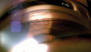
At the slit lamp, with a 78 or 90 diopter lens and a dilated pupil, the examiner sees fibers and sheets in a vitreous that is optically empty in places.
The gel moves freely and collapses easily on eye movement, which is typical of syneresis.
Photopsia occurs when the gel separates from the retina, so flashes after a period of floaters point toward a posterior vitreous detachment (see posterior vitreous detachment).
Differential Diagnosis
- Asteroid hyalosis: small, round, bright calcium soap particles that stay in position in an otherwise intact gel (see asteroid hyalosis)
- Synchysis scintillans: cholesterol crystals that settle with eye movement in a liquefied, usually damaged vitreous (see synchysis scintillans)
- Vitreous hemorrhage: red cells, often with a recent retinal tear or neovascularization (see vitreous hemorrhage)
- Vitritis: inflammatory cells, with anterior chamber reaction and sometimes macular edema
- Retinal tear with pigment cells: tobacco dust in the anterior vitreous, which should prompt a careful peripheral examination
Examination of the Retina
New floaters always call for a dilated examination with scleral depression when the history is suggestive.
The examiner looks for horseshoe tears, operculated holes, retinal dialysis, and lattice degeneration, because all of them can accompany a posterior vitreous detachment (see horseshoe tears).
A patient with isolated, stable, long-standing floaters and a normal retinal periphery has a very low risk of detachment.
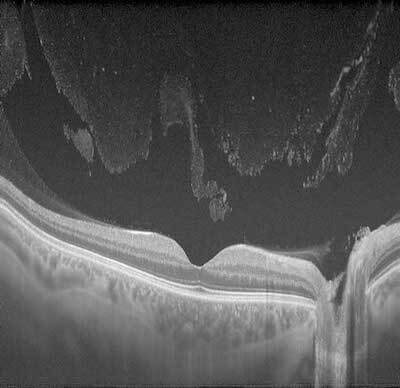
OCT of the macula helps when the patient also complains of distortion, because it can show vitreomacular traction.
Management
Reassurance is the first step, and it is often the only step required.
Explain that the floaters come from the aging gel, that the brain adapts to them over weeks to months, and that most patients stop noticing them.
Warn the patient about the symptoms that need a same-day visit: a sudden shower of new floaters, flashes of light, a dark curtain in the field of vision, or a drop in acuity.
Pars plana vitrectomy removes floaters reliably, but it carries the risks of cataract, retinal tear, and rarely endophthalmitis, so it is reserved for patients whose daily function is clearly reduced (see pars plana vitrectomy).
YAG vitreolysis has been used in selected eyes with a well-defined Weiss ring, but the evidence is limited and results vary.
Nutritional supplements have not been shown to reverse liquefaction.
Syneresis, Posterior Vitreous Detachment, and Later Problems
Liquefaction alone does not harm the retina, but it sets the stage for posterior vitreous detachment, because liquid pockets can break through the posterior cortex and dissect the gel away from the retina.
When the detachment is complete and uneventful, the patient has a new Weiss ring and a period of flashes and floaters that settles over weeks.
When the separation is incomplete, traction at areas of strong adhesion can produce retinal tears, vitreomacular traction, or macular holes.
Understanding this sequence helps the clinician to explain why floaters deserve one careful dilated examination and then, in most patients, no further treatment.


Document what you see
Two smartphone imaging tools built for everyday clinic use — one for the slit lamp, one for the fundus.
From Choroida — the team behind this siteReferences
- Sebag J. Anomalous posterior vitreous detachment: a unifying concept in vitreo-retinal disease. Graefes Arch Clin Exp Ophthalmol. 2004;242:690-698.
- Sebag J. The Vitreous: Structure, Function, and Pathobiology. Springer; 1989.
- Ryan SJ, Schachat AP, Wilkinson CP, et al, eds. Retina. 6th ed. Elsevier; 2018.
- American Academy of Ophthalmology. Basic and Clinical Science Course, Section 12: Retina and Vitreous. AAO; current edition.

Writes and reviews the clinical articles on Educate. Every post is checked against peer-reviewed sources before it goes live.