Conjunctival Kaposi sarcoma is a low-grade vascular tumor of the ocular surface caused by human herpesvirus 8 (HHV-8).
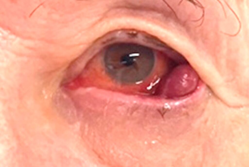
It typically appears as a red to violet patch or nodule on the conjunctiva.
Because it looks like blood under the conjunctiva, patients often receive a diagnosis of subconjunctival hemorrhage first.
That error can delay both tumor care and an HIV diagnosis.
What Causes Conjunctival Kaposi Sarcoma
Chang and colleagues identified HHV-8 DNA sequences in AIDS-associated Kaposi sarcoma in 1994.
The virus drives proliferation of spindle cells and leaky vascular channels.
However, infection alone rarely produces a tumor.
Immune suppression, especially loss of CD4 T cells, allows the lesion to grow.
Four Clinical Forms
Kaposi sarcoma occurs in four recognized settings, and the ocular lesion looks similar in each.
- Classic form in older men of Mediterranean or Eastern European ancestry
- Endemic form in sub-Saharan Africa
- Iatrogenic form after organ transplantation or other immunosuppression
- Epidemic form associated with HIV and AIDS
All-fit Slit-Lamp Adapter
Record and share exactly what you see at the slit lamp. One adapter fits any slit lamp or surgical microscope — and any smartphone.
From Choroida — the team behind this siteThe HIV Clue
In practice, the epidemic form accounts for most ocular presentations.
Therefore, a young adult with a new violaceous conjunctival lesion needs HIV testing.
Also, the eye may show the first visible sign of systemic disease.
Coordinate testing with the primary care or infectious disease team.
Where Conjunctival Kaposi Sarcoma Appears
The inferior fornix and inferior bulbar conjunctiva are frequent sites.
In addition, the eyelid skin and lid margin can develop separate violet nodules.
Lesions may be single or multiple, and both eyes may carry them.
Therefore, evert the lids and examine the fornices in every suspected case.
Slit Lamp Signs
The examiner sees a flat or elevated red-violet lesion with tortuous vessels.
Feeder vessels often run toward the mass.
Nodular lesions feel firm and may bleed after minor trauma.
Conjunctival Kaposi Sarcoma Versus Subconjunctival Hemorrhage
A simple hemorrhage is flat, bright red, and fades over one to three weeks (see subconjunctival hemorrhage).
In contrast, Kaposi sarcoma persists, keeps a purple tone, and often enlarges.
A “hemorrhage” that recurs at the same spot also deserves suspicion.
Then, photograph the lesion and review the patient within a few weeks.
Other Differential Diagnoses
- Pyogenic granuloma or conjunctival granuloma
- Conjunctival lymphoma, which looks salmon-pink
- Amelanotic melanoma
- Capillary hemangioma or varix
- Bacillary angiomatosis in patients with HIV
Dugel Staging
Dugel and colleagues proposed a clinicopathologic staging for adnexal lesions in AIDS.
Stages I and II are flat, patchy lesions under 3 mm high, present for less than four months.
Stage III lesions are nodular, taller than 3 mm, and present longer.
As a result, the stage hints at histology and helps guide treatment.
Biopsy in Conjunctival Kaposi Sarcoma
Tissue confirms the diagnosis when the picture is unclear.
Histology shows spindle cells, slit-like vascular spaces and extravasated red cells.
Also, immunostaining for HHV-8 latency-associated nuclear antigen supports the diagnosis.
However, biopsy can bleed briskly, so plan hemostasis in advance.
Systemic Workup
Every patient needs a full skin and oral examination.
Next, check HIV status, CD4 count and viral load where relevant.
Visceral disease may coexist, so refer to oncology for staging.
Antiretroviral Therapy First
Effective antiretroviral therapy restores immune function and often shrinks Kaposi sarcoma.
Therefore, small, asymptomatic lesions may only need observation once therapy starts.
Similarly, reducing immunosuppression can help iatrogenic disease.
However, regression is slow, and some lesions persist.
Local Treatment of Conjunctival Kaposi Sarcoma
- Surgical excision for small, well-defined nodules
- Cryotherapy for flat lesions
- Intralesional chemotherapy or interferon
- Low-dose external beam radiotherapy for larger or multifocal lesions
Each option carries risks, including scarring, symblepharon, dry eye and recurrence.
Systemic Chemotherapy
Widespread or visceral disease calls for systemic treatment under oncology.
For example, liposomal anthracyclines and taxanes are standard agents for advanced disease.
Ocular lesions usually respond along with the systemic disease.
Meanwhile, the ophthalmologist watches for surface toxicity and residual tumor.
Counseling Patients With Conjunctival Kaposi Sarcoma
Explain that the lesion grows slowly and rarely threatens vision directly.
However, it signals a systemic condition that needs medical attention.
Discuss HIV testing sensitively and with clear consent.
Also, warn that lesions can recur after successful treatment.
Follow-Up of Conjunctival Kaposi Sarcoma
Photograph each lesion at baseline and at every visit.
Then, compare size, color and elevation over time.
New lesions may reflect falling immune control, so inform the treating team.
Pitfalls to Avoid
- Do not label a persistent purple lesion as hemorrhage without follow-up
- Never skip lid eversion and fornix examination
- Avoid aggressive excision before systemic assessment
- Remember that a normal skin exam does not exclude visceral disease


Document what you see
Two smartphone imaging tools built for everyday clinic use — one for the slit lamp, one for the fundus.
From Choroida — the team behind this siteReferences
- Chang Y, Cesarman E, Pessin MS, et al. Identification of herpesvirus-like DNA sequences in AIDS-associated Kaposi’s sarcoma. Science. 1994;266:1865-1869.
- Dugel PU, Gill PS, Frangieh GT, Rao NA. Ocular adnexal Kaposi’s sarcoma in acquired immunodeficiency syndrome. Am J Ophthalmol. 1990;110:500-503.
- Shuler JD, Holland GN, Miles SA, Miller BJ, Grossman I. Kaposi sarcoma of the conjunctiva and eyelids associated with the acquired immunodeficiency syndrome. Arch Ophthalmol. 1989;107:858-862.
- Shields CL, Shields JA. Tumors of the conjunctiva and cornea. Surv Ophthalmol. 2004;49:3-24.
- Bowling B. Kanski’s Clinical Ophthalmology: A Systematic Approach. 8th ed. Elsevier; 2016.

Writes and reviews the clinical articles on Educate. Every post is checked against peer-reviewed sources before it goes live.