
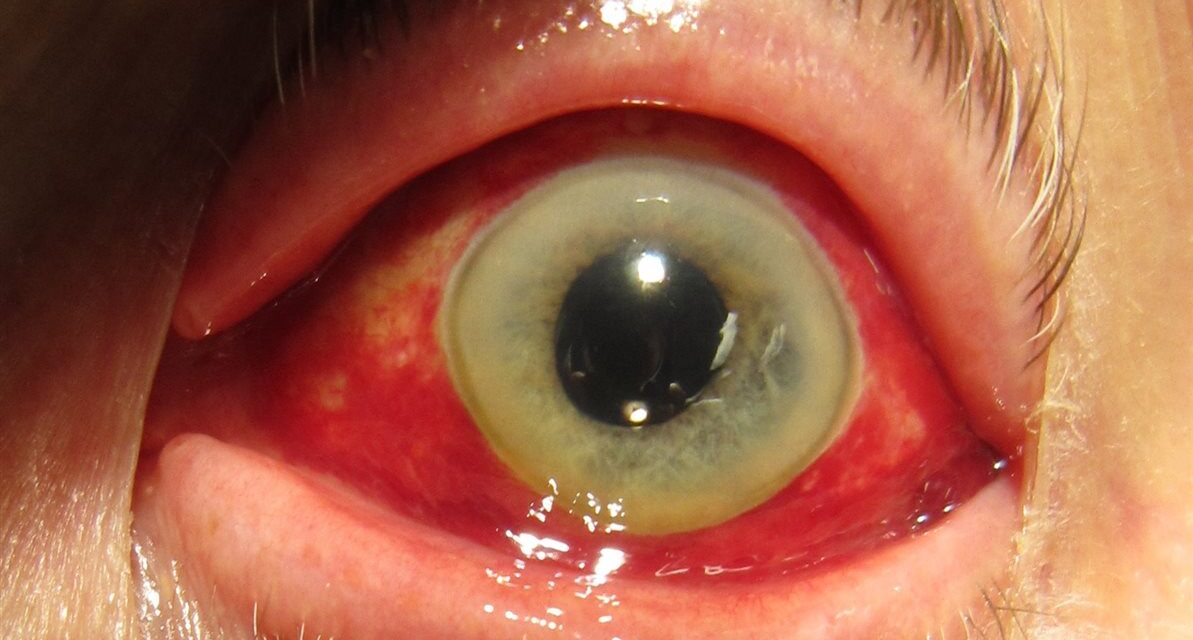
Few ocular findings look more frightening to a patient than a bright red eye that appeared overnight.
A subconjunctival hemorrhage (SCH) is exactly that — a pool of blood trapped beneath the thin, transparent conjunctiva.
It is one of the most common reasons for an urgent, worried phone call to an eye clinic.
The alarming appearance is almost always disproportionate to the actual clinical significance.
In the overwhelming majority of cases, SCH is a benign, self-limited condition that needs nothing more than reassurance.
The clinician’s real job is to quickly separate that majority from the small number of cases that signal trauma, a bleeding disorder, or another condition needing further workup.
What Is Subconjunctival Hemorrhage?
A subconjunctival hemorrhage is bleeding from small conjunctival or episcleral vessels into the potential space beneath the conjunctiva.
Defining features include:
- Sharply demarcated or diffuse bright red blood visible on the white of the eye
- No pain, and typically no change in vision
- A clear cornea and normal pupil, distinguishing it from more serious causes of a red eye
- Blood confined to the subconjunctival space, not extending into the eye itself
The appearance can range from a small, localized patch to blood covering the entire visible sclera, as seen in more extensive cases.
Epidemiology
SCH is extremely common across all age groups and eye care settings.
- It is one of the most frequent causes of an acute, painless red eye presenting for urgent evaluation
- Spontaneous cases increase in frequency with age, likely reflecting more fragile conjunctival vessels and a higher prevalence of hypertension and anticoagulant use
- Many cases go entirely unreported, since patients without pain or vision change may never seek care
Because it is usually self-limited, the true population incidence is almost certainly higher than clinic-based estimates suggest.
All-fit Slit-Lamp Adapter
Record and share exactly what you see at the slit lamp. One adapter fits any slit lamp or surgical microscope — and any smartphone.
From Choroida — the team behind this sitePathophysiology
The conjunctiva and episclera contain a fine network of small, superficial blood vessels.
Bleeding results from rupture of one or more of these vessels, typically triggered by:
- A sudden rise in venous pressure — from coughing, sneezing, vomiting, straining, or heavy lifting
- Direct or blunt trauma, including vigorous eye rubbing
- Vessel fragility related to age or hypertension
- Impaired clotting from anticoagulant or antiplatelet medication, or an underlying bleeding disorder
Once blood escapes into the subconjunctival space, it simply spreads under the transparent conjunctiva and is gradually resorbed, following the same color changes seen in a bruise elsewhere on the body.
Risk Factors
Local and Mechanical Risk Factors
- Valsalva-type events — coughing, sneezing, vomiting, straining, or heavy lifting
- Ocular or periocular trauma, including vigorous rubbing
- Recent ocular surgery or contact lens-related trauma
- Viral conjunctivitis, particularly hemorrhagic conjunctivitis from certain enteroviruses
Systemic Risk Factors
- Systemic hypertension
- Anticoagulant or antiplatelet therapy (warfarin, direct oral anticoagulants, aspirin, clopidogrel)
- Bleeding disorders or coagulopathy
- Diabetes mellitus, related to vascular fragility
A significant proportion of cases remain idiopathic even after a careful history, particularly in otherwise healthy patients.
Clinical Presentation
Symptoms
- Sudden appearance of a red patch on the white of the eye, often first noticed by someone else or in a mirror
- No pain, though a mild foreign-body sensation or gritty feeling is occasionally reported
- No change in visual acuity
- No discharge, distinguishing it from infectious causes of a red eye
Examination Findings
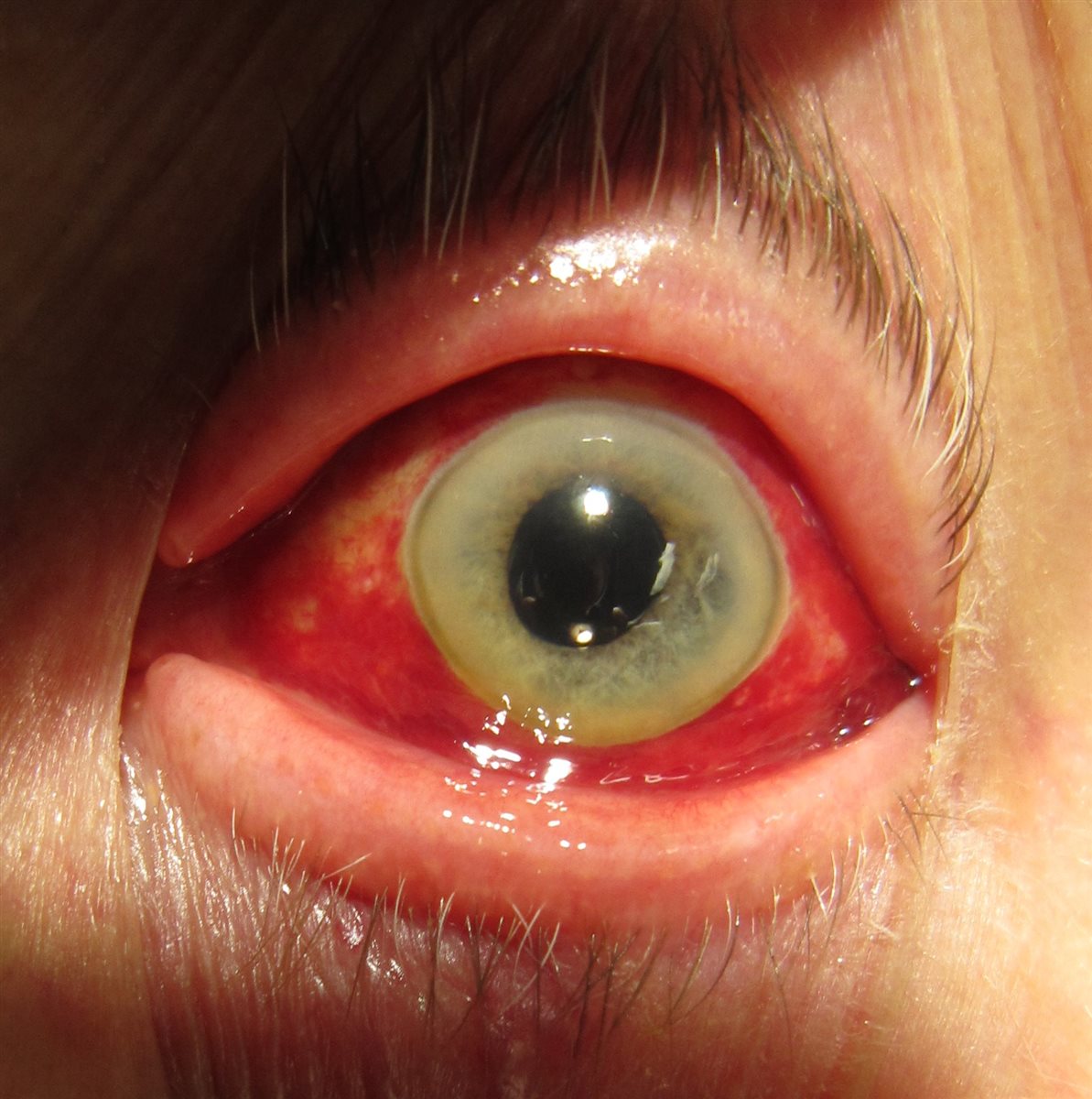
- Flat, bright red blood beneath the conjunctiva, sharply demarcated at the limbus in localized cases or extending diffusely in more extensive ones
- A clear cornea with normal fluorescein staining
- A round, reactive pupil and normal anterior chamber
- Normal visual acuity and intraocular pressure
- No proptosis, restricted eye movement, or lid swelling — findings that, if present, suggest a very different and more urgent diagnosis
A completely normal cornea, pupil, and eye movements alongside the hemorrhage is what confirms this is an isolated, benign finding rather than a marker of orbital or globe injury.
Diagnostic Evaluation
History and Examination
- A focused history covering trauma, Valsalva triggers, anticoagulant use, and bleeding tendency is usually sufficient
- Visual acuity, pupil reactivity, and extraocular movements should be checked and documented as normal
- Blood pressure measurement is reasonable, particularly in patients with recurrent or bilateral hemorrhages
When Further Workup Is Warranted
- Recurrent or bilateral spontaneous hemorrhages without an obvious trigger — consider a coagulation workup
- A clear history of significant trauma — evaluate for globe rupture or orbital fracture rather than assuming isolated SCH
- Hemorrhage with pain, decreased vision, or an abnormal pupil — these findings point away from simple SCH and require full ocular examination
Laboratory testing and imaging are the exception, not the rule — most patients need nothing beyond a careful history and a normal eye exam.
Differential Diagnosis
Conditions that can be confused with or coexist with subconjunctival hemorrhage include:
- Viral or bacterial conjunctivitis — discharge and diffuse conjunctival injection rather than a flat pool of blood
- Episcleritis or scleritis — localized redness with vessel blanching on phenylephrine testing, and often pain in scleritis
- Ruptured globe — history of significant trauma, with an irregular pupil, hyphema, or reduced vision
- Orbital hemorrhage — associated with proptosis, restricted movement, or pain, unlike isolated SCH
- Conjunctival hemangioma or vascular malformation — a longstanding, non-acute lesion rather than a sudden new finding
Ruling out trauma and orbital involvement is the single most important differential step, since these carry very different urgency.
Management
Reassurance and Observation
Appropriate for the vast majority of spontaneous, uncomplicated cases:
- Explain that the hemorrhage will resolve on its own, typically over one to two weeks
- The blood changes color as it resorbs, similar to a bruise, before fully clearing
- Artificial tears can be offered for mild associated irritation
Addressing Underlying Causes
- Optimize blood pressure control if hypertension is identified
- Review anticoagulant or antiplatelet therapy with the prescribing physician if recurrent hemorrhages raise concern — do not stop these medications unilaterally
- Refer for hematologic evaluation if a bleeding disorder is suspected based on recurrent, unprovoked, or bilateral hemorrhages
When Referral Is Needed
- Any hemorrhage associated with trauma significant enough to raise concern for globe rupture
- Hemorrhage with pain, vision change, or an abnormal pupil
- Recurrent hemorrhages without a clear precipitating cause
The overwhelming majority of patients need only reassurance — over-investigation of an isolated, uncomplicated SCH creates unnecessary anxiety and cost.
Prognosis
Prognosis is excellent in essentially all uncomplicated cases.
- Complete resolution is expected within one to two weeks without any lasting effect on vision
- Recurrence can occur, particularly in patients with uncontrolled hypertension or ongoing anticoagulant use
- Vision-threatening outcomes are essentially limited to the rare cases where SCH is actually a marker of trauma or a coexisting bleeding disorder, rather than the hemorrhage itself
Setting expectations early — that this looks worse than it is and will clear on its own — resolves most of the anxiety that brings patients in.
Would you like to document anterior segment findings with your smartphone?


Document what you see
Two smartphone imaging tools built for everyday clinic use — one for the slit lamp, one for the fundus.
From Choroida — the team behind this siteReferences
- Tarlan B, Kiratli H. Subconjunctival hemorrhage: risk factors and potential indicators. Clinical Ophthalmology. 2013.
- Mimura T, Usui T, Yamagami S, et al. Recent causes of subconjunctival hemorrhage. Ophthalmologica. 2010.
- Fukuyama J, Hayasaka S, Yamada K, Setogawa T. Causes of subconjunctival hemorrhage. Ophthalmologica. 1990.
- Subconjunctival Hemorrhage. StatPearls, NCBI Bookshelf.
- Subconjunctival Hemorrhage. EyeWiki, American Academy of Ophthalmology.
Test yourself
A few questions straight from this article.
-
A subconjunctival hemorrhage is a collection of blood in which location?
Blood pools in the potential space beneath the thin transparent conjunctiva and stays confined there, without extending into the eye itself. -
Bleeding in subconjunctival hemorrhage arises from which vessels?
The conjunctiva and episclera carry a fine network of small superficial vessels, and rupture of one or more of these produces the hemorrhage. -
Which everyday event commonly precipitates a spontaneous subconjunctival hemorrhage?
A sudden rise in venous pressure from coughing, sneezing, vomiting, straining or heavy lifting is a classic trigger for vessel rupture. -
Which systemic factor raises the risk of spontaneous subconjunctival hemorrhage?
Impaired clotting from warfarin, direct oral anticoagulants, aspirin or clopidogrel is a recognised systemic risk factor, alongside hypertension and bleeding disorders. -
Which examination profile is expected in an uncomplicated subconjunctival hemorrhage?
A normal cornea, round reactive pupil, normal acuity, pressure and eye movements alongside the blood is what confirms an isolated benign finding rather than globe or orbital injury. -
Which test helps distinguish episcleritis from a subconjunctival hemorrhage?
Episcleritis shows localised redness with vessel blanching on phenylephrine testing, whereas a hemorrhage is a flat pool of blood that does not blanch. -
Which history makes globe rupture, rather than isolated subconjunctival hemorrhage, a concern?
A clear history of significant trauma calls for evaluation for globe rupture or orbital fracture instead of assuming an isolated benign hemorrhage. -
What is appropriate management for a spontaneous, uncomplicated subconjunctival hemorrhage?
The vast majority need only reassurance and observation, with artificial tears for mild irritation; over-investigation creates unnecessary anxiety and cost. -
Over what period does an uncomplicated subconjunctival hemorrhage typically resolve?
Complete resolution is expected within one to two weeks, with the blood changing colour as it resorbs much like a bruise, and no lasting effect on vision. -
How should anticoagulant therapy be handled in a patient with recurrent subconjunctival hemorrhages?
Recurrent hemorrhages justify reviewing anticoagulant or antiplatelet therapy with the prescribing physician; these medications should not be stopped unilaterally in the eye clinic.
