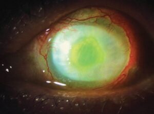
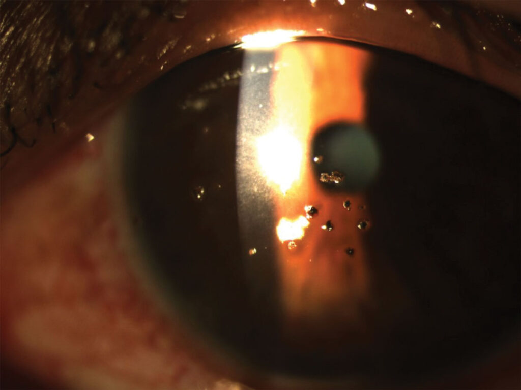
A corneal rust ring is a brown halo of oxidized iron that forms in the corneal stroma around a retained ferrous foreign body.
It is a common finding in workers who grind or weld without eye protection, and in patients whom a metal fragment struck.
The ring itself can be left for a short time, but it should be removed, because it delays healing and can leave a scar.
How a Corneal Rust Ring Forms
When a piece of iron or steel embeds in the cornea, it begins to oxidize within hours because of the moisture in the tissue.
The iron diffuses into the surrounding stroma and stains it.
The stained area becomes a ring of rust around the foreign body.
Within one to two days, a defined ring is visible even if the foreign body has been removed.
The rust is toxic to the keratocytes, and it can impair epithelial healing and cause a local inflammatory reaction.
Other metals, such as copper, can cause a more severe inflammatory reaction.
They need different handling (see corneal foreign body).
Evaluation for Corneal Rust Ring
- Ask about the mechanism, the activity, whether the patient wore safety glasses, and the delay since the injury
- Check the visual acuity, instill a topical anesthetic, and examine with the slit lamp
- Look for the foreign body, the depth, the surrounding infiltrate, and any anterior chamber reaction
- Evert the lids and look for additional foreign bodies in the conjunctival fornices
- Perform a Seidel test with fluorescein if the foreign body could be full thickness
- Dilate the pupil and examine the posterior segment if a high-velocity injury is possible, and obtain imaging for an intraocular foreign body
A history of hammering metal on metal, or a high-speed projectile, calls for imaging of the orbit with CT to exclude an intraocular foreign body.
Check the tetanus immunization status in a patient with a penetrating injury.
All-fit Slit-Lamp Adapter
Record and share exactly what you see at the slit lamp. One adapter fits any slit lamp or surgical microscope — and any smartphone.
From Choroida — the team behind this siteRemoval Technique
- Anesthetize the cornea with topical proparacaine or tetracaine, and use a slit lamp for stability and magnification
- Remove the foreign body first, using a sterile 25-gauge needle bevel up, or a spud
- Remove the rust ring with an Alger brush, a burr at low speed, or the tip of a needle, taking care to stay in the superficial stroma
- Do not aim to remove all the discoloration in one session if it is deep or central, because aggressive removal can scar the cornea
- Leave a small amount of deep rust, which often clears over days to weeks as the keratocytes remove it
Some clinicians remove a central ring on the next day, because the ring softens and detaches more easily after 24 hours.
Protect the patient in the meantime.
Avoid deep drilling, and stop if the stroma becomes thin.
After Removal
Treat the epithelial defect as a corneal abrasion, with a topical antibiotic, and with a cycloplegic if there is significant discomfort or an anterior chamber reaction (see corneal abrasion).
Clinicians do not recommend patching for simple abrasions, and it may increase the risk of infection.
Advise on the use of lubricants and oral analgesics.
Review the patient at 24 to 48 hours to confirm healing, and to look for infection or residual rust.
Topical steroids are rarely needed, and they should be avoided until the epithelium has healed.
Complications of Corneal Rust Ring
- Infectious keratitis, particularly after delayed presentation
- A residual rust stain, which usually clears
- Stromal scarring, especially after deep or aggressive removal
- Recurrent corneal erosion after a large abrasion (see recurrent corneal erosion)
Patients should return promptly if pain, redness, or reduced vision worsens.
Prevention
Emphasize safety eyewear for grinding, welding, hammering, and gardening with power tools.
A short explanation of how easily a small fragment becomes embedded is often more convincing than a general warning.
Special Situations
A rust ring that lies in the visual axis, or a deep ring close to Descemet membrane, should be handled with particular care, and referral to a cornea specialist is reasonable.
Patients who return after several days with an infiltrate and a rust ring need cultures before treatment, because infection can start around a retained foreign body.
In children, sedation may be needed, and a parent should supervise follow-up.
Do not forget to look at the posterior segment in a patient with a history of high-velocity injury.
Documenting the Case
Record the size and depth of the ring, the number of passes needed, and the post-removal appearance.
Photographs are helpful if there is a later claim related to work, since many of these injuries are occupational.
For the latest studies, see the PubMed results for corneal rust ring.


Document what you see
Two smartphone imaging tools built for everyday clinic use — one for the slit lamp, one for the fundus.
From Choroida — the team behind this siteReferences
- Wipperman JL, Dorsch JN. Evaluation and management of corneal abrasions. Am Fam Physician. 2013;87:114-120.
- Mannis MJ, Holland EJ, eds. Cornea. 4th ed. Elsevier; 2017.
- Salmon JF. Kanski’s Clinical Ophthalmology: A Systematic Approach. 9th ed. Elsevier; 2020.
- American Academy of Ophthalmology. Basic and Clinical Science Course, Section 8: External Disease and Cornea. AAO; current edition.

Writes and reviews the clinical articles on Educate. Every post is checked against peer-reviewed sources before it goes live.