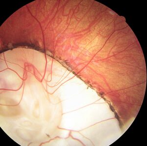
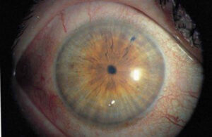
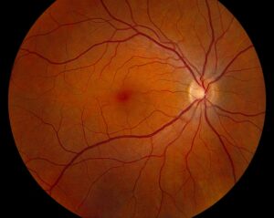
Corectopia is displacement of the pupil away from its normal central position.
A normal pupil sits slightly inferonasal, so mild asymmetry needs no workup.
However, obvious displacement points to disease of the iris, angle or lens.
Therefore, treat corectopia as a sign that needs an explanation.
What Counts as Corectopia
Corectopia describes a pupil displaced toward the iris periphery.
The pupil may stay round or become oval and peaked.
In addition, polycoria often accompanies it in developmental disease.
Congenital Versus Acquired Corectopia
Onset gives the first clue, and old photographs help date it.
For example, a displaced pupil in an infant suggests anterior segment dysgenesis.
On the other hand, new displacement in an adult suggests ICE syndrome, trauma, surgery or inflammation.
All-fit Slit-Lamp Adapter
Record and share exactly what you see at the slit lamp. One adapter fits any slit lamp or surgical microscope — and any smartphone.
From Choroida — the team behind this siteAxenfeld-Rieger Spectrum
Axenfeld-Rieger syndrome combines posterior embryotoxon, iris strands to the cornea and iris hypoplasia.
The pupil often points toward prominent peripheral attachments.
Also, about half of patients develop glaucoma, often in childhood or early adulthood.
Systemic features include dental anomalies and redundant periumbilical skin (see Axenfeld-Rieger syndrome).
Ectopia Lentis et Pupillae
This autosomal recessive disorder displaces the pupil and lens, usually in opposite directions.
Mutations in ADAMTSL4 account for many reported families.
Because both eyes are usually involved, examine the fellow eye carefully.
Other Developmental Causes
- Iris coloboma with an inferonasal keyhole pupil
- Partial aniridia with an eccentric remnant pupil
- Peters anomaly with iridocorneal adhesions
- Isolated congenital corectopia, a diagnosis of exclusion
ICE Syndrome and Acquired Corectopia
ICE syndrome usually affects one eye of a young or middle-aged woman.
Abnormal endothelium migrates across the angle and iris, then contracts.
As a result, the pupil drifts toward the quadrant with the broadest peripheral anterior synechiae.
Progressive iris atrophy shows the most marked displacement, with holes opposite the drift (see ICE syndrome).
Trauma and Corectopia
Blunt injury can tear the sphincter and leave notches in the pupil margin.
Iridodialysis also distorts the pupil, often into a D shape.
Therefore, look for angle recession, phacodonesis and residual hyphema.
Glaucoma after angle recession can appear years later.
Surgical Causes of Corectopia
After cataract surgery, vitreous or iris caught in the wound peaks the pupil toward the incision.
In addition, intraocular lens capture can displace the pupil margin.
Synechiae after a shallow chamber may also pull the pupil.
A peaked pupil with a vitreous strand warrants a check for cystoid macular edema.
Inflammation and Synechiae
Uveitis creates posterior synechiae that bind the pupil margin to the lens.
As a result, the pupil dilates unevenly, which is most obvious after mydriasis.
Look for keratic precipitates, cells and iris atrophy to confirm past inflammation.
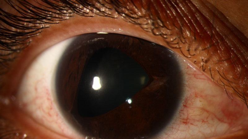
Transient Pupil Distortion
Some pupils distort for minutes and then return to normal.
For example, tadpole pupil reflects segmental spasm of the iris dilator (see tadpole pupil).
Because attacks are brief, patient photographs are valuable.
History in Corectopia
Ask when the change appeared and whether it is progressing.
Then ask about injury, eye surgery, uveitis and family eye disease.
Also, ask about glare, monocular diplopia and dental anomalies in children.
Slit Lamp Sequence
First, assess pupil shape, direction of displacement and the iris surface.
Next, inspect the endothelium for a beaten-metal appearance or posterior embryotoxon.
Then use retroillumination to show iris holes and transillumination defects.
Finally, record pupil reactions and test for a relative afferent pupillary defect.
Gonioscopy and IOP in Corectopia
Gonioscopy maps synechiae, iris strands and angle recession.
Because most causes carry glaucoma risk, measure IOP and examine the optic disc.
Repeat both regularly, even when the pupil looks stable.
Imaging and Additional Tests
Specular microscopy can show ICE cells and support the diagnosis.
Anterior segment OCT or ultrasound biomicroscopy shows angle anatomy and lens position.
Serial photography then documents any progression.
In addition, genetic testing for PITX2, FOXC1 or ADAMTSL4 helps family counseling.
Visual Impact of Corectopia
Many patients with mild displacement see normally.
However, a pupil near the limbus can cause glare and aberrations.
In young children, an eccentric visual axis also risks amblyopia, so refract them under cycloplegia.
Managing Corectopia
Treatment targets the cause and its complications, especially glaucoma.
For example, tinted or artificial iris contact lenses can reduce glare.
For symptomatic eyes, pupilloplasty or an iris prosthesis can recenter the aperture.
However, surgery risks inflammation, bleeding and pressure rise, while ICE membranes may still progress.
Explain to patients that a recentered pupil does not cure the underlying disease.
Pitfalls to Avoid
- Do not label slight inferonasal decentration as disease
- Never skip gonioscopy in a displaced pupil
- Do not confuse ICE syndrome with Axenfeld-Rieger syndrome
- Always check the fellow eye for subtle signs


Document what you see
Two smartphone imaging tools built for everyday clinic use — one for the slit lamp, one for the fundus.
From Choroida — the team behind this siteReferences
- Shields MB, Buckley E, Klintworth GK, Thresher R. Axenfeld-Rieger syndrome: a spectrum of developmental disorders. Surv Ophthalmol. 1985;29:387-409.
- Sacchetti M, Mantelli F, Marenco M, Macchi I, Ambrosio O, Rama P. Diagnosis and management of iridocorneal endothelial syndrome. Biomed Res Int. 2015;2015:763093.
- Christensen AE, Fiskerstrand T, Knappskog PM, Boman H, Rødahl E. A novel ADAMTSL4 mutation in autosomal recessive ectopia lentis et pupillae. Invest Ophthalmol Vis Sci. 2010;51:6369-6373.
- American Academy of Ophthalmology. Basic and Clinical Science Course, Section 10: Glaucoma. San Francisco: AAO; 2023.
- Salmon JF. Kanski’s Clinical Ophthalmology: A Systematic Approach. 9th ed. Edinburgh: Elsevier; 2020.
Test yourself
A few questions straight from this article.
-
Why does slight inferonasal decentration of the pupil not need a corectopia workup?
A normal pupil sits slightly inferonasal, so mild asymmetry needs no workup, while obvious displacement points to disease of the iris, angle or lens. -
A displaced pupil is found in an infant. Which category of cause does this corectopia suggest?
Onset gives the first clue: a displaced pupil in an infant suggests anterior segment dysgenesis, whereas new displacement in an adult suggests ICE syndrome, trauma, surgery or inflammation. -
About what proportion of patients with Axenfeld-Rieger syndrome and corectopia develop glaucoma?
About half of patients with Axenfeld-Rieger syndrome develop glaucoma, often in childhood or early adulthood, alongside posterior embryotoxon, iris strands and iris hypoplasia. -
In ectopia lentis et pupillae, how are the pupil and lens typically displaced?
This autosomal recessive disorder, often from ADAMTSL4 mutations, displaces pupil and lens usually in opposite directions and typically involves both eyes. -
In iridocorneal endothelial (ICE) syndrome, toward which area does the corectopic pupil drift?
Abnormal endothelium migrates across angle and iris and contracts, so the pupil drifts toward the broadest peripheral anterior synechiae, with iris holes opposite the drift in progressive iris atrophy. -
Which test can show ICE cells and support iridocorneal endothelial syndrome as the cause of corectopia?
Specular microscopy can show ICE cells and support the diagnosis, complementing a beaten-metal endothelium seen at the slit lamp. -
After blunt trauma, which injury typically distorts the pupil into a D shape?
Iridodialysis distorts the pupil, often into a D shape, while sphincter tears leave notches in the pupil margin. -
After cataract surgery, a pupil is peaked toward the incision with a vitreous strand. What should be checked?
Vitreous or iris caught in the wound peaks the pupil toward the incision, and a peaked pupil with a vitreous strand warrants a check for cystoid macular edema. -
Why must gonioscopy, IOP and optic disc examination be repeated in a patient with corectopia?
Because most causes of corectopia carry glaucoma risk, gonioscopy maps synechiae and angle recession, and IOP and disc are rechecked regularly even when the pupil looks stable. -
In a young child with corectopia, why is cycloplegic refraction important?
In young children an eccentric visual axis risks amblyopia, so they are refracted under cycloplegia; a pupil near the limbus can also cause glare and aberrations.

Writes and reviews the clinical articles on Educate. Every post is checked against peer-reviewed sources before it goes live.