Foveal schisis is a splitting of the neurosensory retina at the fovea, usually without leakage on angiography.
Clinicians often call the sign a spoke-wheel macula because of its radial folds.
However, several diseases produce it, and OCT helps separate them.
What Foveal Schisis Looks Like
Fundus examination shows fine radial striae around the foveal center.
The foveal reflex may also look dull or absent.
Patients usually report slow central blur or difficulty reading.
In contrast, sudden loss suggests a complication, such as a macular hole, hemorrhage or detachment.
Why the Spoke-Wheel Pattern Forms
Henle fibers radiate obliquely from the fovea.
Therefore, cavities in this region often take a stellate or petaloid outline.
Bridging tissue between the cavities creates the visible spokes.
The underlying mechanism varies, because adhesion defects, traction and possible drug effects can all contribute.
Fundus Explorer Pro
Photograph the retinal findings described here with the phone already in your pocket — 22 D optics and built-in illumination in one handheld unit.
From Choroida — the team behind this siteHistory That Narrows the Foveal Schisis Differential
Start with age, sex, refraction, family history and medications.
For example, a boy with bilateral reading difficulty and affected maternal male relatives suggests X-linked disease.
High myopia, on the other hand, points toward traction.
Next, ask directly about high-dose niacin and taxane chemotherapy, since patients may not mention them.
Reading the OCT Layers
First, identify the layer that contains the cavities.
Then, inspect the vitreous, internal limiting membrane (ILM), epiretinal membrane and ellipsoid zone.
Also look for subretinal fluid, a staphyloma and any connection to the optic disc.
However, layer location alone rarely proves the cause, because patterns overlap between diseases.
X-Linked Foveal Schisis
Pathogenic RS1 variants disrupt retinoschisin, a protein involved in retinal cell adhesion.
OCT often shows cavities in the inner nuclear layer, although other layers may also split.
Some patients also have peripheral schisis, so examine the periphery carefully.
With age, the cavities may flatten and leave atrophy (see X-linked retinoschisis).
Confirming and Treating X-Linked Disease
Electroretinography often shows a reduced b-wave, but a normal result does not exclude the diagnosis.
Therefore, RS1 testing provides the confirmation.
Topical dorzolamide reduced cystic spaces in some patients in a small study, although visual gain was limited.
Gene therapy remains investigational, so patients should hear that no approved option exists.
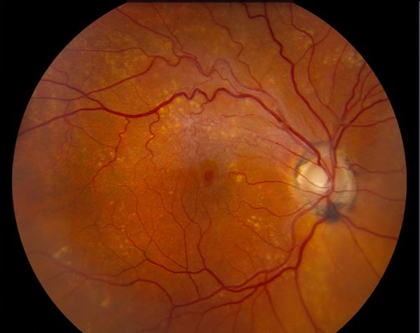
Myopic Foveal Schisis
Takano and Kishi described this finding in severely myopic eyes with posterior staphyloma.
OCT commonly shows outer retinal splitting, sometimes with inner cavities as well.
Vitreous traction, epiretinal membrane, a stiff ILM and the staphyloma may all contribute.
As a result, the sign belongs within the wider spectrum of myopic traction maculopathy.
Managing the Myopic Eye
Stable eyes with good vision can usually remain under observation.
However, progressive vision loss or foveal detachment may justify vitrectomy with ILM peeling.
Fovea-sparing peeling aims to lower the risk of postoperative macular hole, but the supporting evidence comes mainly from surgical series.
Surgery also cannot reverse established atrophy, so counsel patients about limited visual recovery.
Foveal Schisis Without Leakage or Family History
Stellate nonhereditary idiopathic foveomacular retinoschisis (SNIFR) enters the differential here.
Ober and colleagues described stellate splitting in the outer plexiform layer without angiographic leakage.
Because SNIFR is a diagnosis of exclusion, first rule out traction, drugs, optic pit and inherited disease.
Drug-Related Foveal Schisis
Gass described cystoid maculopathy without typical leakage in patients taking high-dose nicotinic acid.
Taxanes can also cause cystoid spaces with little or no angiographic leakage.
Next, discuss any drug change with the prescribing physician, especially during cancer treatment.
Optic Pit and Peripapillary Disease
Always examine the disc before settling on a macular diagnosis.
An optic pit can produce schisis-like cavities that track from the disc margin, sometimes with subretinal fluid.
Therefore, extend the OCT scan through the optic nerve head.
Separating Foveal Schisis From Macular Edema
Diabetes, vein occlusion, uveitis and surgery can all cause cystoid macular edema.
Fluorescein angiography typically shows petaloid leakage in these conditions (see leakage patterns).
In contrast, schisis usually shows little or no leakage.
As a result, anti-VEGF agents and steroids have a weaker rationale when leakage is absent.
Follow-Up Planning in Foveal Schisis
Repeat OCT with the same protocol, because small scan shifts can mimic change.
At each visit, compare cavity height, foveal contour, ellipsoid zone integrity and subretinal fluid.
In myopic eyes, new metamorphopsia should prompt earlier review.
Pitfalls to Avoid
- Do not label every radial macular pattern as X-linked disease
- Do not treat schisis as edema before checking angiography when doubt exists
- Avoid missing an optic pit by scanning only the fovea
- Do not rely on OCT improvement as proof of visual benefit


Document what you see
Two smartphone imaging tools built for everyday clinic use — one for the slit lamp, one for the fundus.
From Choroida — the team behind this siteReferences
- Takano M, Kishi S. Foveal retinoschisis and retinal detachment in severely myopic eyes with posterior staphyloma. Am J Ophthalmol. 1999;128:472-476.
- Ober MD, Freund KB, Shah M, et al. Stellate nonhereditary idiopathic foveomacular retinoschisis. Ophthalmology. 2014;121:1406-1413.
- Apushkin MA, Fishman GA. Use of dorzolamide for patients with X-linked retinoschisis. Retina. 2006;26:741-745.
- Shimada N, Sugamoto Y, Ogawa M, et al. Fovea-sparing internal limiting membrane peeling for myopic traction maculopathy. Am J Ophthalmol. 2012;154:693-701.
- Gass JD. Nicotinic acid maculopathy. Am J Ophthalmol. 1973;76:500-510.

Writes and reviews the clinical articles on Educate. Every post is checked against peer-reviewed sources before it goes live.