Bell’s phenomenon is the upward and usually outward rotation of the eye during forced lid closure.
Charles Bell described it in 1823 while studying facial palsy.
Clinically, it shows how well the cornea can hide when the lids fail to close.
Therefore, this brief test can influence exposure treatment and ptosis surgery planning.
What Bell’s Phenomenon Looks Like
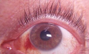
The globe rolls upward as the orbicularis contracts, and the cornea moves beneath the upper lid.
Many eyes also drift slightly outward.
In facial palsy, you can often watch this movement through the open fissure.
Patients rarely report it because the response is involuntary.
Anatomy Behind the Reflex
The facial nerve drives lid closure, while the oculomotor nerve supplies the superior rectus and inferior oblique.
Brainstem connections probably coordinate these systems, although the exact pathway remains uncertain.
Facial palsy weakens closure but usually spares the elevators.
As a result, the upward roll often persists on the paralyzed side.
All-fit Slit-Lamp Adapter
Record and share exactly what you see at the slit lamp. One adapter fits any slit lamp or surgical microscope — and any smartphone.
From Choroida — the team behind this siteBlinks and Forced Closure
Search-coil recordings show that ordinary blinks produce a small downward and nasal eye movement.
By contrast, upward rotation belongs mainly to forced or sustained closure.
Therefore, a strong response cannot replace normal blinking, which spreads the tear film.
Eye position during sleep also varies, so a good daytime test cannot guarantee nighttime protection.
How to Test Bell’s Phenomenon
The examination takes seconds and needs no equipment.
- First, ask the patient to close both eyes firmly.
- Next, gently lift one upper lid while the patient keeps squeezing.
- Then note the direction of rotation and how much cornea remains visible.
- Finally, repeat on the other side and compare.
Grading the Response
No single grading system is universal, so describe what you see.
For example, a good response hides the entire cornea under the upper lid.
A fair response leaves part of the cornea visible, while a poor response leaves it largely exposed.
Also measure lagophthalmos in millimeters during gentle closure, because forced closure can hide the gap.
Normal Variation in Bell’s Phenomenon
Healthy people do not all show a classic upward roll.
Francis and Loughhead documented this variability in 508 patients.
Therefore, an absent response does not prove disease by itself.
However, new asymmetry deserves more attention.
Upgaze Palsy and Bell’s Phenomenon
A preserved upward roll despite poor voluntary elevation suggests a lesion above the oculomotor nuclei.
However, poor effort and restriction can confuse the test.
Therefore, interpret it with ductions and the full motility examination.
Bell’s Phenomenon and Corneal Exposure
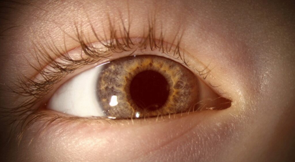
A poor response leaves the cornea in the path of any lagophthalmos.
Therefore, expect earlier inferior or interpalpebral punctate staining.
A good response offers partial protection, yet the inferior cornea can still dry.
Always combine the test with fluorescein staining and corneal sensation testing.
The BAD Eye in Facial Palsy
Clinicians use the BAD mnemonic to flag facial palsy patients at high risk of corneal damage.
- B: absent Bell’s response
- A: corneal anesthesia
- D: dry eye
As a result, these patients may report little pain despite significant surface breakdown.
Managing Exposure When the Response Is Poor
Lubricating drops by day and ointment at night support the surface, but they cannot restore closure.
Taping can add overnight protection, although poor technique may injure the cornea.
Next, consider an upper lid weight or tarsorrhaphy for persistent exposure.
Explain that these measures protect vision, and some may need revision.
Bell’s Phenomenon in Ptosis Surgery
Lifting the upper lid widens the fissure and can worsen lagophthalmos.
Therefore, test the response before any ptosis repair, along with levator function and the tear film.
A poor response generally favors a more conservative lift.
This may leave some residual droop, but it can reduce the risk of exposure keratopathy.
High-Risk Ptosis Patients
Myopathic ptosis deserves caution because orbicularis weakness and limited elevation often coexist.
For example, patients with Kearns-Sayre ptosis may have little corneal protection after surgery.
Third nerve palsy and myasthenia gravis raise similar concerns.
Counseling and Follow-Up in Bell’s Phenomenon
Warn patients about dryness, foreign body sensation and blurred vision after the lid is raised.
Next, plan lubrication before surgery and arrange an early staining check.
Ask them to report pain, redness or falling vision promptly.
At follow-up, repeat lagophthalmos measurement and fluorescein staining.
Pitfalls to Avoid
- Do not treat a good response as complete corneal protection
- Never rely on the test without measuring lagophthalmos
- Do not assume that absence always means pathology
- Do not skip corneal sensation in a patient with facial palsy


Document what you see
Two smartphone imaging tools built for everyday clinic use — one for the slit lamp, one for the fundus.
From Choroida — the team behind this siteReferences
- Bell C. On the motions of the eye, in illustration of the uses of the muscles and nerves of the orbit. Philos Trans R Soc Lond. 1823;113:166-186.
- Francis IC, Loughhead JA. Bell’s phenomenon: a study of 508 patients. Aust J Ophthalmol. 1984;12:15-21.
- Collewijn H, van der Steen J, Steinman RM. Human eye movements associated with blinks and prolonged eyelid closure. J Neurophysiol. 1985;54:11-27.
- Rucker JC. Normal and abnormal lid function. Handb Clin Neurol. 2011;102:403-424.
- American Academy of Ophthalmology. Basic and Clinical Science Course, Section 7: Oculofacial Plastic and Orbital Surgery. San Francisco: AAO.

Writes and reviews the clinical articles on Educate. Every post is checked against peer-reviewed sources before it goes live.