
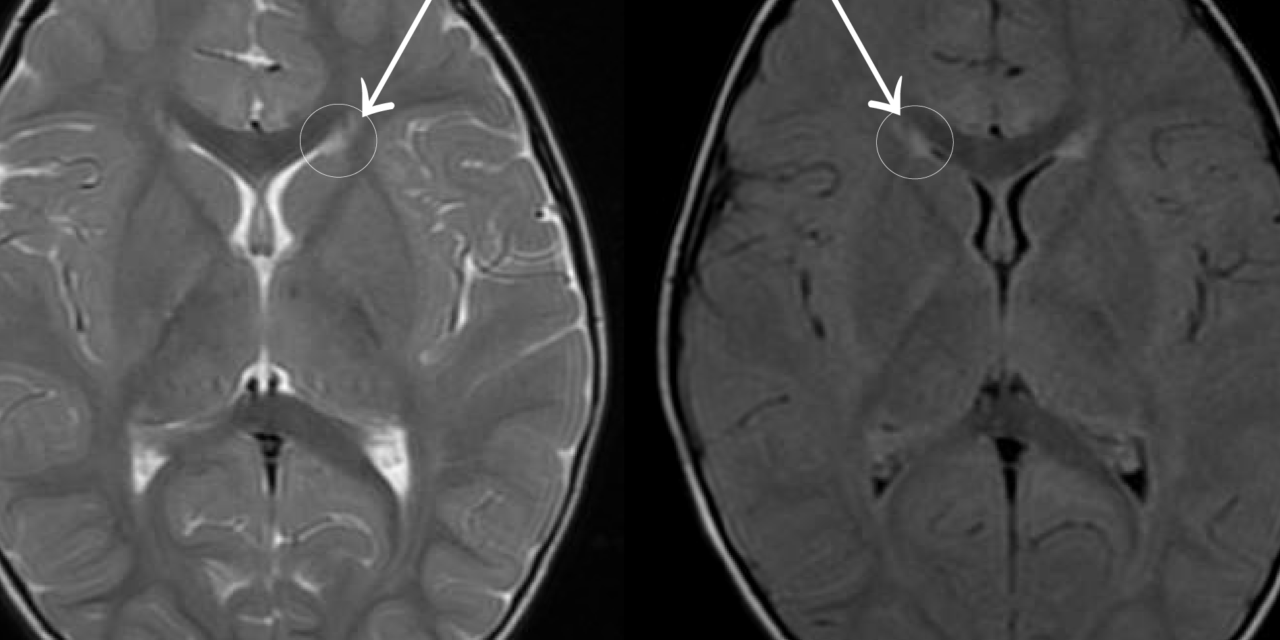
Periventricular leukomalacia is a pattern of white matter injury affecting premature infants, and its visual consequences illustrate an important principle in pediatric neuro-ophthalmology: a child can have entirely normal-appearing eyes on examination and still have profound visual impairment, because the injury lies not in the eye itself but in the white matter pathways carrying and processing visual information within the brain.
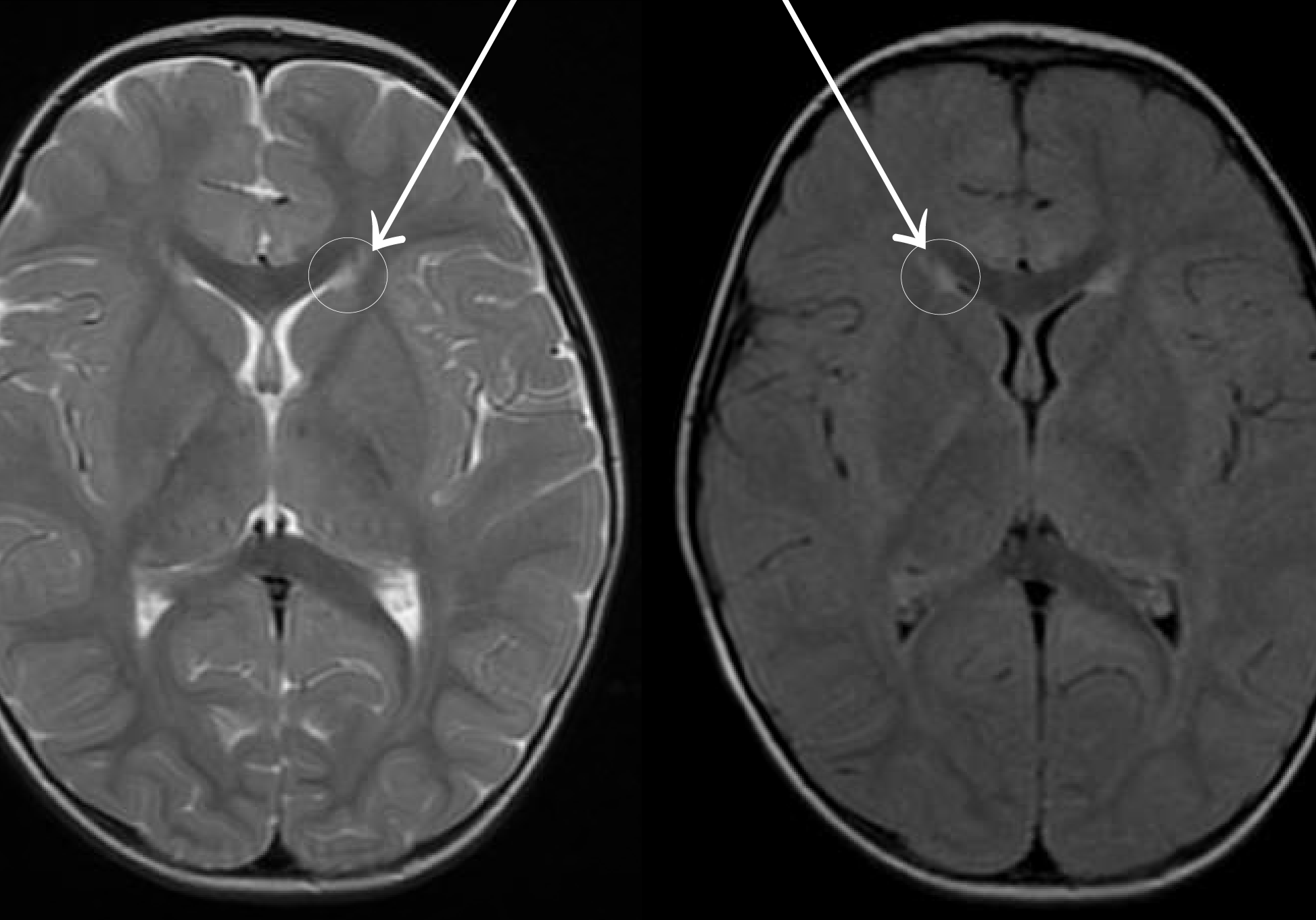
What Periventricular Leukomalacia Is
Periventricular leukomalacia is injury to the white matter surrounding the lateral ventricles of the brain, occurring predominantly in premature infants, most often those born before roughly 32 weeks gestation, and it results from the particular vulnerability of this periventricular white matter to ischemia and inflammation during a specific window of brain development.
The optic radiations, the white matter tracts carrying visual information from the lateral geniculate nucleus to the occipital visual cortex, pass directly through this periventricular region, which is why periventricular leukomalacia is one of the most common causes of cerebral, or cortical, visual impairment in children.
Why the Optic Radiations Are Especially Vulnerable
The optic radiations occupy a substantial portion of the periventricular white matter, and their location places them squarely within the zone most commonly affected by the watershed ischemic injury pattern characteristic of periventricular leukomalacia.
This anatomic vulnerability explains the strong, well-documented association between periventricular leukomalacia and visual impairment, even when other neurological consequences of the same brain injury, such as spastic diplegia, may be less severe or absent.
Clinical Presentation
- Reduced visual behavior in infancy, including poor visual attention, reduced fixation and following, and inconsistent visual responses, often noted before a formal diagnosis of periventricular leukomalacia is confirmed
- A relatively normal-appearing anterior visual examination in many cases: normal pupillary responses, normal-appearing optic discs (though optic atrophy can develop secondarily in more severe cases), and no structural abnormality of the eye itself
- Visual field defects, often affecting the inferior visual field disproportionately, reflecting the specific location of optic radiation fibers most commonly damaged
- Frequently accompanied by other neurodevelopmental findings associated with prematurity and periventricular white matter injury, including motor impairment
- A characteristic, though not universal, pattern of visual behavior in cerebral visual impairment more broadly: difficulty with visual crowding, preference for certain colors or moving stimuli, and inconsistency in visual responses across different settings and times of day
Diagnosis
- A history of prematurity with known or suspected periventricular leukomalacia, often first identified on neonatal cranial ultrasound or later confirmed with MRI
- Ophthalmic examination to exclude an ocular cause for the reduced vision, since a normal eye examination in the setting of a known history of periventricular leukomalacia supports a cerebral origin for the visual impairment
- Formal visual function assessment, including behavioral visual acuity testing appropriate to the child’s developmental level, and visual field assessment when feasible
- MRI findings characteristic of periventricular leukomalacia, including periventricular white matter loss and ventricular enlargement, particularly involving the trigone region where optic radiation fibers are concentrated, correlate with visual outcome
Management
There is no treatment that reverses the underlying white matter injury, so management centers on maximizing functional visual use and supporting overall development.
- Early involvement of vision rehabilitation services, including teachers of the visually impaired and low vision specialists experienced with cerebral visual impairment
- Environmental adaptations that account for the specific visual processing difficulties common in cerebral visual impairment, such as reducing visual clutter and allowing extra time for visual processing
- Coordinated care with neurodevelopmental and physical therapy services, given the frequent coexistence of motor and other developmental needs
- Correction of any coexisting refractive error or ocular finding, such as strabismus, which is common in this population and can be addressed even though it does not address the underlying cortical component
Prognosis
Visual outcome in periventricular leukomalacia varies widely, correlating with the extent and location of white matter injury seen on imaging, and ranges from mild visual processing difficulty to severe visual impairment.
Some degree of visual improvement over the early years of life is common in cerebral visual impairment broadly, reflecting both neuroplasticity and improved developmental visual strategies, which supports early and sustained engagement with vision-specific rehabilitation services even when initial visual function is significantly impaired.


Document what you see
Two smartphone imaging tools built for everyday clinic use — one for the slit lamp, one for the fundus.
From Choroida — the team behind this siteReferences
- Jacobson L, Ek U, Fernell E, Flodmark O, Broberger U. Visual impairment in preterm children with periventricular leukomalacia–visual, cognitive and neuropaediatric characteristics related to cerebral imaging findings. Dev Med Child Neurol. 1996;38:724-735.
- Fazzi E, Bova SM, Uggetti C, et al. Visual-perceptual impairment in children with periventricular leukomalacia. Brain Dev. 2004;26:506-512.
- Hoyt CS. Visual function in the brain-damaged child. Eye (Lond). 2003;17:369-384.
- Volpe JJ. Brain injury in premature infants: a complex amalgam of destructive and developmental disturbances. Lancet Neurol. 2009;8:110-124.
Test yourself
A few questions straight from this article.
-
Periventricular leukomalacia occurs predominantly in infants born before roughly which gestational age?
Periventricular leukomalacia affects premature infants, most often those born before roughly 32 weeks gestation, when the periventricular white matter is most vulnerable. -
Periventricular leukomalacia is injury to which part of the brain?
It is injury to the white matter around the lateral ventricles, which is particularly vulnerable to ischaemia and inflammation at that stage of brain development. -
Which visual pathway structure passes directly through the region damaged in periventricular leukomalacia?
The optic radiations run through the periventricular white matter, which is why periventricular leukomalacia is among the commonest causes of cerebral visual impairment in children. -
The optic radiations carry visual information between which two structures?
They are the white matter tracts conveying visual information from the lateral geniculate nucleus to the occipital visual cortex. -
A child with visual impairment from periventricular leukomalacia typically shows which ocular examination finding?
Pupillary responses and optic discs usually look normal with no structural eye abnormality, because the injury lies in the brain rather than the eye. -
Visual field defects in periventricular leukomalacia disproportionately affect which part of the field?
Field loss often favours the inferior field, reflecting the specific location of the optic radiation fibres most commonly damaged. -
Which MRI findings in periventricular leukomalacia correlate with visual outcome?
Periventricular white matter loss and ventricular enlargement, particularly at the trigone where optic radiation fibres concentrate, correlate with visual outcome. -
Which pattern of visual behaviour is characteristic of cerebral visual impairment?
Children often struggle with visual crowding, prefer certain colours or moving stimuli, and respond inconsistently across different settings and times of day. -
Management of visual impairment from periventricular leukomalacia centres on which aim?
No treatment reverses the underlying white matter injury, so care focuses on functional vision, rehabilitation and supporting overall development. -
What visual course is common over the early years of life in cerebral visual impairment?
Some improvement is common, reflecting neuroplasticity and better developmental visual strategies, which supports early and sustained rehabilitation engagement.
