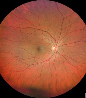
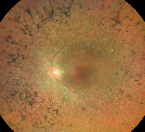
Retinitis pigmentosa sine pigmento is a form of retinitis pigmentosa in which the fundus shows little or no bone-spicule pigmentation, even though the functional loss is that of a rod-cone dystrophy.
It is easy to overlook, because a normal-looking retina in a patient with night blindness invites other diagnoses.
The electroretinogram is the key to the diagnosis, so request it early too.
What Is Retinitis Pigmentosa Sine Pigmento?
The term applies to eyes with typical symptoms and ERG changes of retinitis pigmentosa, but without the visible pigment migration of the classic disease.
Doctors also generally regard it as an early or atypical stage of rod-cone dystrophy and not a separate entity.
Pigment may appear later as the disease progresses.
Some patients show attenuated vessels, mild pigment epithelial changes, or a waxy pallor of the disc only.
In addition, the same genes that cause classic retinitis pigmentosa can lead to this presentation.
The pattern of inheritance can be autosomal dominant, autosomal recessive, or X-linked (see retinitis pigmentosa).
Clinical Features of Retinitis Pigmentosa Sine Pigmento
- Night blindness, often the first symptom, with difficulty adapting to darkness
- Progressive loss of the peripheral visual field, with ring scotomas
- Normal or mildly reduced acuity in the early stage
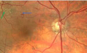
- Normal or near-normal fundus, with possible arteriolar attenuation and mild RPE mottling
- Fine vitreous cells and posterior subcapsular cataract in some cases
- Macular edema, which can reduce acuity (see macular edema causes)
Fundus Explorer Pro
Photograph the retinal findings described here with the phone already in your pocket — 22 D optics and built-in illumination in one handheld unit.
From Choroida — the team behind this siteInvestigations
The full-field ERG shows reduced rod and cone responses, with rod function lost earlier (see electroretinography).
Visual fields show a ring scotoma or a constricted field, and dark adaptometry shows an elevated threshold.
OCT may also show loss of the ellipsoid zone in the periphery and preservation at the center, and fundus autofluorescence may show a ring of increased signal around the fovea.
Genetic testing identifies the gene and the mode of inheritance, and it helps with counselling and with eligibility for trials.
Differential Diagnosis of Retinitis Pigmentosa Sine Pigmento
- Congenital stationary night blindness, which is nonprogressive and has a different ERG pattern (see congenital stationary night blindness)
- Vitamin A deficiency, which causes night blindness that responds to supplementation (see vitamin A deficiency)
- Cancer-associated retinopathy and autoimmune retinopathy (see autoimmune retinopathy)
- Cone-rod dystrophy and other inherited retinal disease (see cone-rod dystrophy)
- Unilateral causes: old inflammation, trauma, or diffuse unilateral subacute neuroretinitis (see unilateral pigmentary retinopathy)
A careful history and the ERG usually settle the diagnosis.
Management for Retinitis Pigmentosa Sine Pigmento
There is no cure, but several measures help.
- Genetic counselling and testing for the patient and relatives
- Low-vision services and orientation and mobility training as the field narrows
- Treatment of macular edema with carbonic anhydrase inhibitors or other options
- Cataract surgery when the lens opacity affects vision
- Sunglasses and light protection for comfort
- Vitamin A palmitate was shown in one trial to slow the loss of ERG amplitude, but its use is individualized and needs medical supervision
Also, gene therapy is available for biallelic RPE65-associated disease and is being studied for others (see gene therapy for inherited retinal disease).
Explain to the patient that the disease progresses slowly in most cases, and that regular examinations help to identify treatable complications.
Why It Is Missed
Doctors often reassure a patient with night blindness and a normal fundus, or think the problem is functional.
The diagnosis may be delayed for years.
A thorough history of night vision, dark adaptation, and peripheral awareness should prompt a full-field ERG even when the retina looks normal.
Children with a family history of night blindness deserve the same approach, since early disease can be subtle.
Genetic Counselling
The family history is essential: ask about night blindness, early blindness, consanguinity, and hearing loss in relatives.
Also, testing by gene panel or exome sequencing identifies the cause in a majority of patients, and the result guides the risk to children and siblings.
Discuss that the same mutation can cause different phenotypes in a family, and that a family member who has no symptoms may still carry the gene.
Follow-Up
Examine patients annually with visual fields, OCT of the macula, and acuity, and consider repeat ERG every few years or when there is a change in symptoms.
Look for complications, such as macular edema, cataract, and epiretinal membrane, since they respond to treatment.
Provide information about patient organizations, research registries, and clinical trials, which many families find helpful.
Practical Tip
When the ERG is reduced but the fundus is normal, ask about medications and nutritional status, and test serum vitamin A, since deficiency is treatable and can mimic the disease.
Document the findings carefully, because a pigment may appear years later and the early notes help to confirm the course.
For the latest studies, see the PubMed results for retinitis pigmentosa sine pigmento.


Document what you see
Two smartphone imaging tools built for everyday clinic use — one for the slit lamp, one for the fundus.
From Choroida — the team behind this siteReferences
- Hartong DT, Berson EL, Dryja TP. Retinitis pigmentosa. Lancet. 2006;368:1795-1809.
- Berson EL. Retinitis pigmentosa: the Friedenwald Lecture. Invest Ophthalmol Vis Sci. 1993;34:1659-1676.
- Ryan SJ, Schachat AP, Wilkinson CP, et al, eds. Retina. 6th ed. Elsevier; 2018.
- American Academy of Ophthalmology. Basic and Clinical Science Course, Section 12: Retina and Vitreous. AAO; current edition.

Writes and reviews the clinical articles on Educate. Every post is checked against peer-reviewed sources before it goes live.