Waxy disc pallor is a pale, yellowish optic nerve appearance classically seen in retinitis pigmentosa (RP).
It belongs to the familiar fundus triad, along with bone spicule pigment and arteriolar attenuation.
However, the sign is neither universal nor specific.
Therefore, it should prompt a search for diffuse retinal disease.
What Waxy Disc Pallor Looks Like
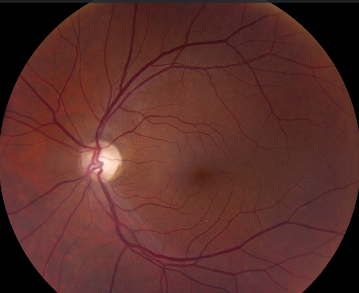
The disc looks smooth, dull and pale yellow, somewhat like candle wax.
Also, the margins usually stay fairly distinct, and the cup is generally not deep.
Both discs tend to look similar because RP is typically bilateral.
Small surface vessels may appear sparse, while the retinal arterioles look narrow.
Why the Disc Looks Waxy
The exact mechanism remains uncertain, although two explanations fit the available evidence.
First, glial tissue can proliferate on or within the nerve head.
This tissue may alter how the disc reflects light, and thus its apparent color.
Second, reduced disc capillary flow may remove some of the normal pink tone.
Fundus Explorer Pro
Photograph the retinal findings described here with the phone already in your pocket — 22 D optics and built-in illumination in one handheld unit.
From Choroida — the team behind this siteThe Retinal Disease Behind the Color
RP begins with photoreceptor dysfunction, usually affecting rods before cones.
As photoreceptors die, the outer retina consumes less oxygen, and this may contribute to vascular attenuation.
Meanwhile, inner retinal neurons survive in greater numbers than photoreceptors, although ganglion cells can also decline.
As a result, the pale disc does not necessarily indicate severe primary optic neuropathy.
Examining the Disc Carefully
The sign usually appears late, so a normal disc color does not exclude early RP.
Start with a dilated view, then compare both discs and assess the cup, rim and vessels.
Also, inspect the mid-peripheral retina for pigment migration and atrophy.
Media can mislead you, because cataract adds yellow while pseudophakia can make a disc look paler.
Waxy Disc Pallor Versus Optic Atrophy
Primary optic atrophy often looks chalky white and may show sectoral or temporal patterns.
In contrast, the waxy disc has a yellow tone and accompanies widespread retinal change.
Glaucoma usually adds rim loss and cupping, whereas compressive disease often causes pallor beyond the amount of cupping.
A relative afferent pupillary defect or poor color vision early in the course should raise concern for optic nerve disease.
Other Causes of Similar Pallor
Several retinal and optic nerve disorders can resemble this appearance.
Therefore, the surrounding fundus and the history guide the differential:
- Cone-rod dystrophy and other inherited retinal dystrophies
- Syndromic RP, including Usher and Bardet-Biedl syndromes
- Autoimmune or cancer-associated retinopathy
- Syphilitic or congenital rubella pseudoretinitis pigmentosa
- Thioridazine toxicity and advanced hydroxychloroquine toxicity
- Compressive lesions, such as optic nerve sheath meningioma
OCT Findings With Waxy Disc Pallor
OCT helps separate outer retinal loss from optic nerve damage.
RP typically shows ellipsoid zone loss outside a central island, with outer nuclear layer thinning.
Peripapillary nerve fiber measurements vary, and some patients show thickening while others show thinning.
Consequently, a thin nerve fiber layer does not alone establish glaucoma or compression.
Function Tests That Confirm the Diagnosis
Full-field electroretinography provides the main functional evidence for generalized rod and cone dysfunction.
In typical RP, rod responses decline early and often more severely than cone responses.
Visual fields then document ring scotoma or constriction, while autofluorescence maps retinal damage.
Finally, genetic testing can refine inheritance, prognosis and eligibility for gene-specific treatment.
Waxy Disc Pallor and Prognosis
Disc color alone offers a poor estimate of visual function.
For that reason, track symptoms, fields, OCT ellipsoid zone width and acuity over time.
Photographs can document appearance, but they do not replace functional testing.
Can Waxy Disc Pallor Reverse?
No treatment is established to restore normal disc color.
Voretigene neparvovec improved functional vision in a phase 3 trial, but only for confirmed biallelic RPE65 retinopathy.
However, it does not suit most RP genotypes and carries surgical and immune-related risks.
Therefore, treatment goals should focus on retinal function, while the disc serves as a clinical clue.
Counseling Patients About Waxy Disc Pallor
Explain that the color reflects a chronic process affecting the retina and optic nerve head.
Next, make clear that the sign does not predict blindness on a fixed timeline.
Patients often benefit from low vision services, mobility advice and genetic counseling.
Finally, look for treatable problems such as cystoid macular edema and cataract, because they can reduce vision further.
Pitfalls to Avoid
- Do not diagnose RP from disc color alone
- Do not assume every pale disc reflects retinal dystrophy
- Avoid calling pallor glaucoma without matching cup and rim changes
- Never skip the pupil and color vision assessment


Document what you see
Two smartphone imaging tools built for everyday clinic use — one for the slit lamp, one for the fundus.
From Choroida — the team behind this siteReferences
- Hartong DT, Berson EL, Dryja TP. Retinitis pigmentosa. Lancet. 2006;368:1795-1809.
- Verbakel SK, van Huet RAC, Boon CJF, et al. Non-syndromic retinitis pigmentosa. Prog Retin Eye Res. 2018;66:157-186.
- Stone JL, Barlow WE, Humayun MS, de Juan E Jr, Milam AH. Morphometric analysis of macular photoreceptors and ganglion cells in retinas with retinitis pigmentosa. Arch Ophthalmol. 1992;110:1634-1639.
- Russell S, Bennett J, Wellman JA, et al. Efficacy and safety of voretigene neparvovec (AAV2-hRPE65v2) in patients with RPE65-mediated inherited retinal dystrophy. Lancet. 2017;390:849-860.

Writes and reviews the clinical articles on Educate. Every post is checked against peer-reviewed sources before it goes live.