Idiopathic CNV is choroidal neovascularization in a patient, usually under 50, with no identifiable ocular or systemic cause.
Because exclusion defines the label, the diagnosis stays provisional until the workup is complete.
Most eyes respond well to anti-VEGF therapy.
What Defines Idiopathic CNV
New vessels grow from the choroid through a break in Bruch membrane into the subretinal space.
In consensus nomenclature, this pattern corresponds to type 2 macular neovascularization (see MNV types).
However, drusen, high myopia, lacquer cracks, chorioretinal scars and angioid streaks should all be absent.
Who Gets Idiopathic CNV
Patients are usually healthy young or middle-aged adults.
In Cohen’s series of young patients, myopia was the leading cause of CNV.
As a result, every new case deserves a deliberate search for a more common explanation.
Fundus Explorer Pro
Photograph the retinal findings described here with the phone already in your pocket — 22 D optics and built-in illumination in one handheld unit.
From Choroida — the team behind this siteSymptoms and Fundus Signs in Idiopathic CNV
Patients report sudden central blur, metamorphopsia or a small central scotoma.
Pain and redness are absent, so their presence should prompt a search for inflammation.
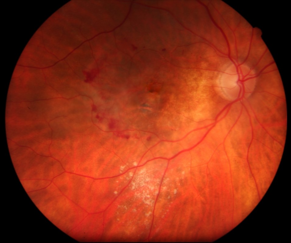
The typical lesion is a small gray-green subretinal membrane near the fovea.
A rim of subretinal hemorrhage often surrounds it, which explains the sudden symptoms.
Next, inspect the whole fundus for punched-out scars, peripapillary atrophy and angioid streaks.
OCT in Idiopathic CNV
OCT usually shows hyperreflective subretinal material above the RPE, with adjacent subretinal fluid.
Intraretinal fluid may also appear, although it is often modest.
In addition, drusen or pigment epithelial detachments suggest another disease.
Angiography and OCTA
Fluorescein angiography classically shows early lacy hyperfluorescence, followed by late leakage.
Blood may block part of the signal, so margins can look smaller than the true lesion.
OCT angiography then displays the network without dye, which suits repeat visits.
However, hemorrhage can hide flow, so a negative scan needs cautious interpretation.
Mimics on Imaging
Several lesions can resemble active neovascularization.
For example, vitelliform material, focal choroidal excavation and chronic central serous chorioretinopathy can show subretinal fluid.
Therefore, combine OCT, OCTA and dye studies before committing a patient to repeated injections.
Causes to Exclude in Idiopathic CNV
Consider these conditions before accepting the label:
- High myopia, with lacquer cracks or staphyloma (see myopic CNV)
- Punctate inner choroidopathy and multifocal choroiditis
- Presumed ocular histoplasmosis and old toxoplasmosis scars
- Angioid streaks and traumatic choroidal rupture
- Best disease and pachychoroid neovasculopathy
Targeted Workup for Idiopathic CNV
Ask about refractive surgery, blunt trauma, photopsia, uveitis and tuberculosis or histoplasmosis exposure.
Then obtain cycloplegic refraction, axial length, dilated examination and fundus autofluorescence.
Laboratory testing should follow the clinical findings, because no single panel fits every patient.
Syphilis serology and tuberculosis testing are reasonable when history raises suspicion.
Also, vitritis or vascular sheathing should redirect the evaluation (see retinal vasculitis).
Natural History of Idiopathic CNV
Ho and colleagues described a more favorable course in untreated idiopathic subfoveal lesions than in neovascular AMD.
Some membranes regress and leave a small scar.
However, others cause permanent central damage, and outcome cannot be predicted at presentation.
Anti-VEGF Treatment for Idiopathic CNV
Anti-VEGF agents reduce vascular leakage and suppress membrane activity.
MINERVA, a randomized sham-controlled trial, showed better two-month acuity with ranibizumab in CNV of uncommon causes, including idiopathic cases.
Mandal’s bevacizumab series also reported anatomical and visual improvement.
However, these drugs do not eliminate the membrane or repair established scarring.
Endophthalmitis and intraocular inflammation are rare but serious risks, so patients need clear warning symptoms.
Injection Schedule and Other Options
Many clinicians give one injection, then reassess at about four weeks.
Further doses follow only for persistent fluid, hemorrhage or new symptoms.
Thermal laser and photodynamic therapy now have limited roles because of scarring risk.
Inflammatory disease may also need corticosteroids alongside anti-VEGF therapy.
Counseling Patients with Idiopathic CNV
Explain that idiopathic means no cause has been found so far.
Discuss realistic goals, because distortion can persist despite a dry OCT.
Women planning pregnancy should also discuss the limited safety data for intravitreal anti-VEGF.
Follow-Up in Idiopathic CNV
Review monthly until the lesion is inactive, then lengthen intervals gradually.
Teach daily monocular Amsler grid checks, because recurrence can present with new distortion.
Finally, examine the fellow eye at each visit.
Pitfalls to Avoid
- Do not label a lesion idiopathic before checking refraction and axial length
- Do not overlook small peripheral punched-out scars
- Never rely on a single imaging modality for a suspicious lesion
- Avoid broad laboratory panels without clinical direction


Document what you see
Two smartphone imaging tools built for everyday clinic use — one for the slit lamp, one for the fundus.
From Choroida — the team behind this siteReferences
- Ho AC, Yannuzzi LA, Pisicano K, DeRosa J. The natural history of idiopathic subfoveal choroidal neovascularization. Ophthalmology. 1995;102:782-789.
- Cohen SY, Laroche A, Leguen Y, Soubrane G, Coscas GJ. Etiology of choroidal neovascularization in young patients. Ophthalmology. 1996;103:1241-1244.
- Lai TYY, Staurenghi G, Lanzetta P, et al. Efficacy and safety of ranibizumab for the treatment of choroidal neovascularization due to uncommon causes: twelve-month results of the MINERVA study. Retina. 2018;38:1464-1477.
- Mandal S, Garg S, Venkatesh P, et al. Intravitreal bevacizumab for subfoveal idiopathic choroidal neovascularization. Arch Ophthalmol. 2007;125:1487-1492.
- Spaide RF, Jaffe GJ, Sarraf D, et al. Consensus nomenclature for reporting neovascular age-related macular degeneration data. Ophthalmology. 2020;127:616-636.

Writes and reviews the clinical articles on Educate. Every post is checked against peer-reviewed sources before it goes live.