Retinal hemorrhage recovery usually takes weeks to months, depending on the layer involved and the underlying cause.
Small intraretinal hemorrhages may clear within weeks, while thick subretinal blood may leave permanent damage.
Also, a cleared fundus does not guarantee restored vision.
Retinal Hemorrhage Recovery: Typical Timelines
Published data on clearance times are limited, so these ranges are clinical estimates.
- Flame hemorrhages: often weeks
- Dot-blot hemorrhages: weeks to several months
- Subhyaloid hemorrhages: weeks to months
- Vitreous hemorrhage: weeks to many months
- Subretinal hemorrhage: often months, with variable scarring
Why the Layer Sets the Clock
First, blood location determines how readily macrophages and retinal vessels can clear it.
Depth also determines which tissue the blood damages.
Therefore, begin by identifying the layer (see hemorrhage patterns by layer).
Fundus Explorer Pro
Photograph the retinal findings described here with the phone already in your pocket — 22 D optics and built-in illumination in one handheld unit.
From Choroida — the team behind this siteFlame and Dot-Blot Hemorrhages
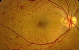
Flame hemorrhages lie in the nerve fiber layer and often resorb fairly quickly.
Dot-blot hemorrhages sit deeper in the middle retina and may persist longer.
However, new lesions keep appearing while the vascular disease stays active.
Subhyaloid Blood and Retinal Hemorrhage Recovery
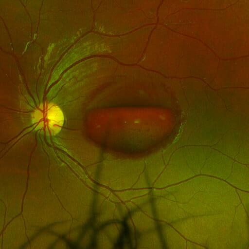
Preretinal blood collects beneath the hyaloid or internal limiting membrane, often forming a boat shape.
Because this space clears poorly, a dense premacular pool may linger for weeks or months.
Vision often improves after clearance if the macula remains healthy.
Vitreous Hemorrhage Clearing Time
Red cells disperse through the gel, then clear slowly.
Thin hemorrhage may settle within weeks, whereas dense hemorrhage may persist for months.
Next, a dense hemorrhage without a fundus view requires B-scan ultrasound to exclude retinal detachment or a tear.
Submacular Hemorrhage Carries the Worst Prognosis
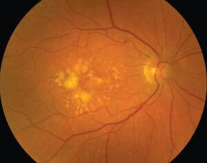
Subretinal blood separates photoreceptors from the retinal pigment epithelium, which supports them.
In addition, experimental work implicates fibrin, clot retraction and iron toxicity in early outer retinal injury.
Therefore, thick foveal blood may damage vision before the clot clears.
Retinal Hemorrhage Recovery by Cause
Cause determines whether bleeding is a single event or a recurring process.
For example, a Valsalva hemorrhage usually reflects one brief pressure spike.
In contrast, diabetic retinopathy and vein occlusion may keep bleeding until treatment controls the disease.
Diabetes and Retinal Vein Occlusion
Diabetic hemorrhages often fluctuate with retinopathy severity and systemic control.
After vein occlusion, hemorrhages commonly fade over months while edema and nonperfusion determine vision (see branch retinal vein occlusion).
Therefore, track macular thickness and perfusion as well as the visible blood.
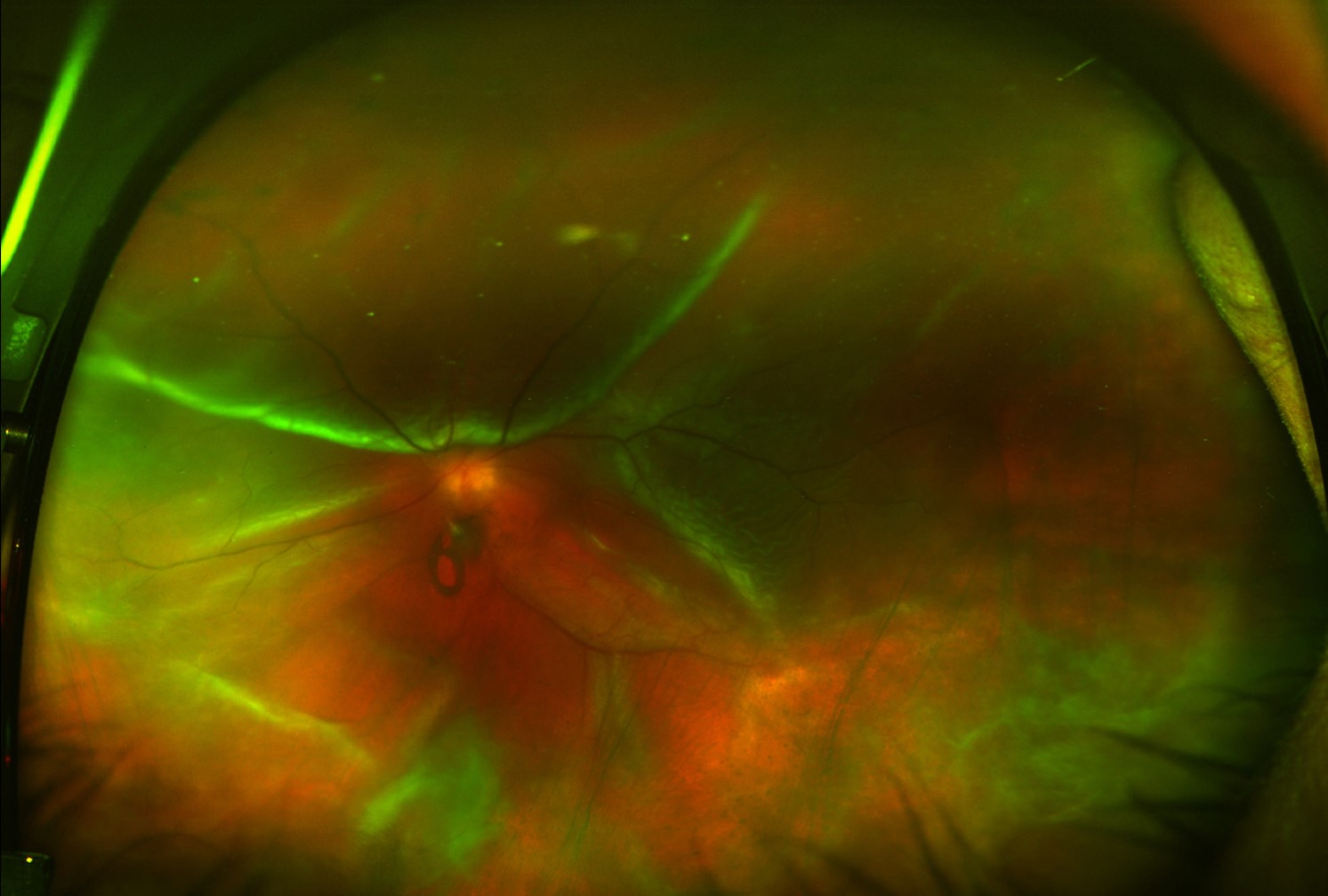
Newborn, Valsalva and Altitude Hemorrhages
Most newborn hemorrhages clear within days to a few weeks (see newborn hemorrhages).
Valsalva hemorrhages often resolve spontaneously, although dense premacular blood may take longer.
Finally, atypical patterns or unexplained persistence should prompt a broader evaluation.
When Vision Is Unlikely to Recover
Blood obscures vision, but structural damage sets the final limit.
These findings suggest a guarded prognosis:
- Thick subfoveal hemorrhage or subretinal fibrosis
- Macular ischemia on angiography
- Disrupted ellipsoid zone after clearance
- Chronic macular edema with inner retinal disorganization
Imaging During Retinal Hemorrhage Recovery
OCT localizes blood and shows the underlying outer retina, although dense hemorrhage can block the signal.
Serial scans then reveal whether the ellipsoid zone returns as the shadow fades.
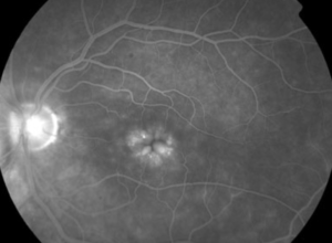
Fluorescein angiography helps identify ischemia, but blood may mask fluorescence.
Treatments That May Shorten Retinal Hemorrhage Recovery
Observation suits many small hemorrhages, particularly when the fovea is spared.
Nd:YAG hyaloidotomy can drain selected premacular hemorrhages, although it carries risks including macular hole.
For diabetic vitreous hemorrhage, vitrectomy clears the media quickly.
However, DRCR Protocol AB found no significant difference in mean acuity over 24 weeks versus initial aflibercept.
Submacular hemorrhage may warrant vitrectomy, tissue plasminogen activator or pneumatic displacement, yet these options cannot reverse established photoreceptor loss.
Examination Pearls During Retinal Hemorrhage Recovery
First, document hemorrhage size and location with a drawing or fundus photograph at each visit.
Then compare these records to judge whether the blood is shrinking or spreading.
Also, a color change from red to yellow-green suggests hemoglobin breakdown and resolution.
However, an enlarging hemorrhage should prompt a search for an active bleeding source.
Lesions That Mimic Hemorrhage
However, choroidal nevus, melanoma and retinal arterial macroaneurysm can resemble a hemorrhage.
Therefore, confirm the diagnosis with imaging when the blood does not clear as expected.
Follow-Up and Counseling
Patients should report new floaters, flashes, a curtain or sudden vision loss immediately.
Dense vitreous hemorrhage needs repeat ultrasound until the retina becomes visible.
Also, give patients a range, because exact clearing dates are unreliable.
Finally, separate two questions clearly: when the blood will clear and how much vision will return.
Pitfalls to Avoid
- Never promise full vision because the hemorrhage looks small
- Always perform B-scan when dense blood hides the retina
- Check for hypertension, diabetes or blood disorders


Document what you see
Two smartphone imaging tools built for everyday clinic use — one for the slit lamp, one for the fundus.
From Choroida — the team behind this siteReferences
- Toth CA, Morse LS, Hjelmeland LM, Landers MB 3rd. Fibrin directs early retinal damage after experimental subretinal hemorrhage. Arch Ophthalmol. 1991;109:723-729.
- Avery RL, Fekrat S, Hawkins BS, Bressler NM. Natural history of subfoveal subretinal hemorrhage in age-related macular degeneration. Retina. 1996;16:183-189.
- Central Vein Occlusion Study Group. Natural history and clinical management of central retinal vein occlusion. Arch Ophthalmol. 1997;115:486-491.
- Emerson MV, Pieramici DJ, Stoessel KM, et al. Incidence and rate of disappearance of retinal hemorrhage in newborns. Ophthalmology. 2001;108:36-39.
- Glassman AR, Beaulieu WT, Maguire MG, et al. Effect of initial intravitreous anti-vascular endothelial growth factor therapy vs vitrectomy for vitreous hemorrhage. JAMA. 2020;324:2383-2395.

Writes and reviews the clinical articles on Educate. Every post is checked against peer-reviewed sources before it goes live.