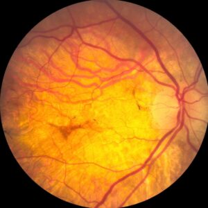
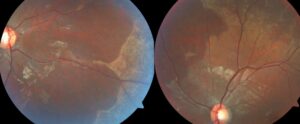
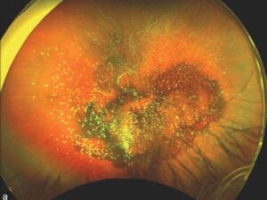
Gunn dots are tiny, glistening white specks seen on the normal inner retinal surface.
They usually appear around the optic disc and along the major vessel arcades.
Because they sparkle during ophthalmoscopy, trainees may mistake them for crystals or exudates.
However, they have no established pathological significance and need no treatment.
What Gunn Dots Are
Robert Marcus Gunn described these reflective points in the nineteenth century.
Most authors attribute them to light reflecting from the internal limiting membrane, possibly near Müller cell footplates.
However, published histologic and imaging data are limited.
Therefore, this explanation is best viewed as a reasonable anatomical model.
Where to Look for Gunn Dots
Start at the optic disc margin, then follow the superior and inferior arcades.
The dots often lie beside large vessels and scatter across the posterior pole.
Both eyes commonly show a similar pattern, although the number may differ slightly.
Marked asymmetry should prompt a closer look, especially when other retinal changes accompany it.
Fundus Explorer Pro
Photograph the retinal findings described here with the phone already in your pocket — 22 D optics and built-in illumination in one handheld unit.
From Choroida — the team behind this siteWhat the Examiner Sees
Each dot looks small, discrete, and white or yellow-white, with a crisp glint.
The surrounding retina keeps its normal color and transparency.
Also, you should find no edema, hemorrhage, pigment change, or vascular abnormality nearby.
Those negative findings support the diagnosis as much as the dots themselves.
Why the Sparkle Shifts
A surface reflex depends on the angle between the light beam and the retina.
Therefore, some dots may brighten or vanish as you tilt the lens or move the slit beam.
By contrast, a lesion within the retina usually keeps a more stable appearance.
This simple maneuver takes seconds and often settles the question at the slit lamp.
Examining Gunn Dots at the Slit Lamp
First, use a dilated pupil and a 78 D or 90 D lens.
Next, narrow the beam and vary its angle across the peripapillary retina.
Red-free light can then enhance the contrast of inner retinal reflexes.
Indirect ophthalmoscopy offers a wider view, while the slit lamp better shows fine surface detail.
OCT and Autofluorescence
Few studies have characterized this finding with multimodal imaging.
Therefore, no validated OCT signature currently defines it.
In practice, OCT helps most by showing intact retinal layers and no intraretinal fluid.
Similarly, autofluorescence should show no associated lesion pattern (see retinal photography methods).
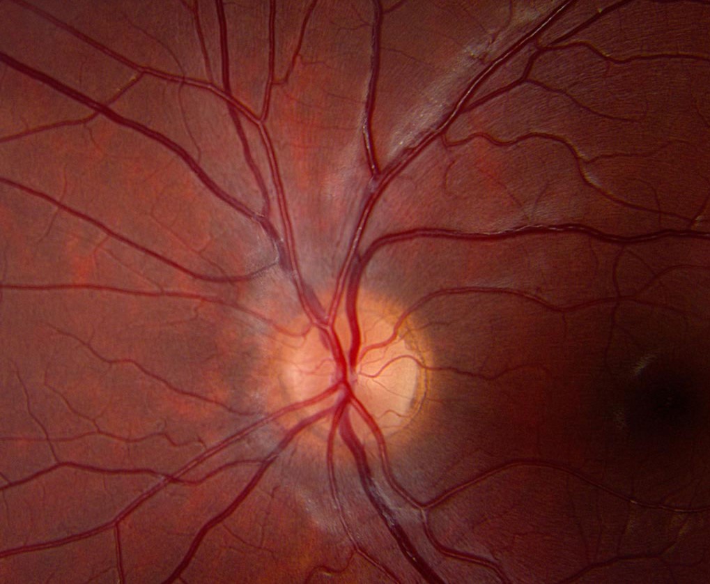
Gunn Dots Versus Crystalline Retinopathy
Crystals may follow tamoxifen, canthaxanthin, talc exposure, or oxalosis.
They also occur in Bietti crystalline dystrophy and macular telangiectasia type 2.
Therefore, ask about medications, intravenous drug use, renal disease, and family history.
Crystals usually persist across viewing angles, and they often accompany atrophy, vascular change, or macular disease.
Alport Flecks and Other Mimics
Alport syndrome can produce dot-and-fleck retinopathy around the macula.
Because these flecks can look subtle, ask about hematuria, renal disease, and hearing loss.
Benign fleck retina and fundus albipunctatus also produce multiple pale lesions.
However, those flecks are not surface glints, and fundus albipunctatus also causes night blindness.
Drusen and Exudates
Drusen sit beneath the retina and look duller, with softer edges.
OCT then shows them at the level of the retinal pigment epithelium (see peripheral drusen).
Hard exudates are yellow and waxy, and they often form rings around leaking vessels.
Moreover, they usually coexist with thickening, microaneurysms, or another vascular cause.
When to Investigate Further
Isolated, symmetric, typical dots in an asymptomatic young patient need no special tests.
However, investigate when vision falls, lesions look atypical, or macular changes coexist.
Also consider further evaluation after high-risk drug exposure or with renal and hearing symptoms.
OCT is a sensible first test, and additional imaging should follow the suspected cause.
Counseling Patients About Gunn Dots
Use plain language and explain that these bright points are a normal light reflection.
Next, reassure the patient that they do not indicate retinal damage or require treatment.
You can also mention that they often become less noticeable with age.
Clear documentation then prevents repeated alarm, especially if another clinician notices them later (see fundus variants).
Pitfalls to Avoid
- Do not label every retinal glint as normal without checking the macula
- Avoid diagnosing Gunn dots when vision loss remains unexplained
- Never skip the medication history when bright specks look crystalline
- Do not order extensive testing for a typical isolated finding
- Avoid judging progression from photographs taken at different angles


Document what you see
Two smartphone imaging tools built for everyday clinic use — one for the slit lamp, one for the fundus.
From Choroida — the team behind this siteReferences
- Drenser K, Sarraf D, Jain A, Small KW. Crystalline retinopathies. Surv Ophthalmol. 2006;51:535-549.
- Nadim F, Walid H, Adib J. The differential diagnosis of crystals in the retina. Int Ophthalmol. 2001;24:113-121.
- Savige J, Ratnaike S, Colville D. Retinal abnormalities characteristic of inherited renal disease. J Am Soc Nephrol. 2011;22:1403-1415.
- American Academy of Ophthalmology. Basic and Clinical Science Course, Section 12: Retina and Vitreous. San Francisco: AAO.
- Sadda SR, Schachat AP, Wilkinson CP, et al. Ryan’s Retina. 7th ed. Philadelphia: Elsevier; 2022.
Test yourself
A few questions straight from this article.
-
Where in the fundus do Gunn dots usually appear?
Gunn dots usually sit around the disc and along the major arcades, often beside large vessels, and they scatter across the posterior pole. -
Most authors attribute the glint of Gunn dots to light reflecting from which structure?
Reflection from the internal limiting membrane, possibly near Müller cell footplates, is the usual explanation, but limited data make it a reasonable model rather than proven fact. -
Which slit lamp maneuver helps show that a bright speck is a Gunn dot?
A surface reflex depends on the angle of the light, so some dots brighten or vanish as the lens tilts or the beam moves, whereas an intraretinal lesion stays more stable. -
Which surrounding finding supports the diagnosis of Gunn dots?
The nearby retina keeps its normal color and transparency, and the absence of edema, hemorrhage, pigment change or vascular abnormality supports the diagnosis as much as the dots do. -
How does OCT help most in practice when Gunn dots are suspected?
No validated OCT signature defines Gunn dots, so the test is useful mainly for showing intact retinal layers without intraretinal fluid. -
Which feature favors crystalline retinopathy over Gunn dots?
Crystals usually persist whatever the viewing angle and often accompany atrophy, vascular change or macular disease, unlike a shifting surface reflex. -
Dot-and-fleck retinopathy around the macula should prompt asking about which symptoms, given its association with Alport syndrome?
Alport syndrome can produce subtle dot-and-fleck retinopathy around the macula, so the history should cover hematuria, renal disease and hearing loss. -
How should isolated, symmetric, typical Gunn dots in an asymptomatic young patient be managed?
This typical picture needs no special tests; investigation is for falling vision, atypical lesions, coexisting macular change, high-risk drug exposure or renal and hearing symptoms. -
On OCT, at what level do drusen lie when they are being separated from Gunn dots?
Drusen sit beneath the retina, look duller with softer edges, and appear on OCT at the level of the retinal pigment epithelium. -
Which appearance identifies hard exudates rather than Gunn dots?
Hard exudates are yellow and waxy, often ring leaking vessels, and usually coexist with thickening, microaneurysms or another vascular cause.

Writes and reviews the clinical articles on Educate. Every post is checked against peer-reviewed sources before it goes live.