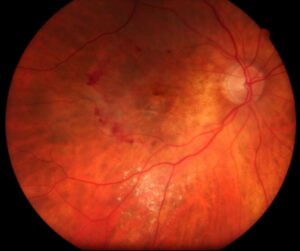
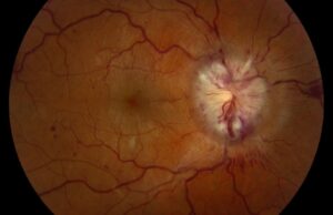
Temporal disc pallor is whitening of the temporal optic disc rim, and it can signal papillomacular bundle loss.
However, the temporal rim normally looks paler than the nasal rim.
Therefore, examiners should confirm the sign with visual function testing and imaging.
What Temporal Disc Pallor Means
Axons from the macula enter the disc mainly on its temporal side.
When these axons degenerate, that sector loses neural tissue and small surface vessels.
As a result, the rim looks white or chalky.
The sign marks established damage, but it does not name the cause (see optic atrophy).
Why the Papillomacular Bundle Is Vulnerable
Small-caliber axons dominate this bundle, and they may tolerate energy failure poorly.
For example, pathology studies in Leber hereditary optic neuropathy show preferential loss of smaller axons.
Therefore, patients often notice reduced acuity and color vision first.
Fundus Explorer Pro
Photograph the retinal findings described here with the phone already in your pocket — 22 D optics and built-in illumination in one handheld unit.
From Choroida — the team behind this siteTrue Temporal Disc Pallor or Normal Variation?
Healthy discs usually have a thinner, paler temporal rim.
In addition, a large physiologic cup can expose the pale lamina on that side.
Symmetry, normal color vision and normal imaging all favor a normal variant.
By contrast, asymmetry, reduced function and matching tissue loss support true disease.
Examining Temporal Disc Pallor
First, use the same lens and light setting for both eyes.
Next, compare the temporal rims directly and look for loss of fine surface capillaries.
Red-free light can then reveal nerve fiber layer defects between the disc and macula.
Finally, check for a relative afferent pupillary defect, although symmetric disease may not produce one.
Symptoms and Function Tests
Patients may report blurred central vision, faded colors or difficulty reading.
Color plates and red desaturation can reveal dysfunction when acuity remains fairly good.
Central or cecocentral scotomas fit papillomacular disease.
OCT in Temporal Disc Pallor
OCT can confirm whether the pale sector has truly lost tissue.
Temporal peripapillary thinning and macular ganglion cell layer loss support the diagnosis.
However, normative databases perform less reliably in high myopia and tilted discs.
Inherited Causes of Temporal Disc Pallor
Dominant optic atrophy often shows a wedge-shaped temporal pallor, sometimes with temporal excavation.
It typically causes slow, bilateral central vision loss beginning in childhood.
Leber hereditary optic neuropathy, by contrast, usually causes subacute, sequential loss in young adults.
Family history therefore helps, although a negative history does not exclude either condition.
Toxic and Nutritional Causes
Ethambutol, methanol and heavy alcohol or tobacco exposure can all damage central axons (see methanol toxicity).
Vitamin B12 and folate deficiency can produce a similar bilateral picture.
Early discs may look normal, and pallor may appear only later.
Therefore, take a detailed medication, diet and substance history.
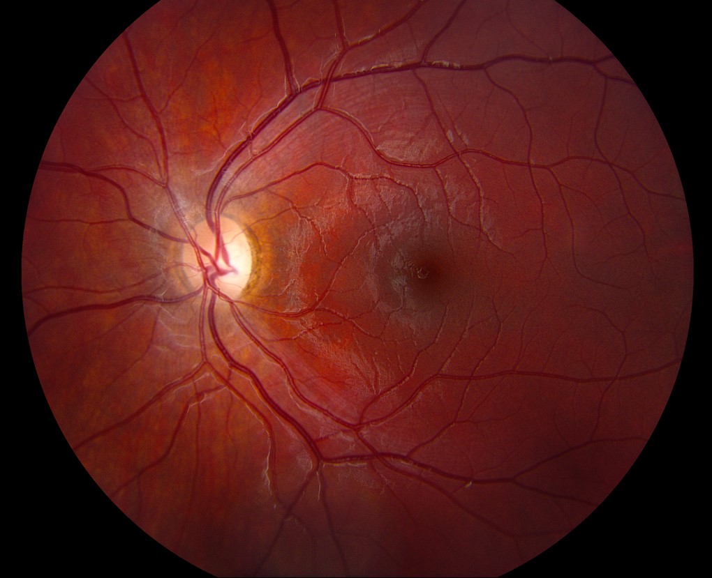
Demyelination and Temporal Pallor
Temporal pallor commonly follows optic neuritis, especially in patients with multiple sclerosis.
The history usually includes earlier painful vision loss, followed by partial recovery.
However, some patients recall no clear attack.
Compressive Lesions Can Look Similar
Compression usually causes progressive visual loss, and its pallor may be diffuse or sectoral.
Chiasmal lesions can produce band atrophy, with bitemporal field loss.
Therefore, unexplained progression or a field defect respecting the vertical meridian calls for neuroimaging.
Temporal Disc Pallor Versus Glaucoma
Glaucoma characteriztically produces cupping with a preserved rim color.
In contrast, nonglaucomatous neuropathy often shows rim pallor that exceeds the degree of cupping.
Greenfield and colleagues linked younger age, reduced acuity, vertically aligned field loss and such pallor to intracranial lesions.
Workup for Temporal Disc Pallor
Testing should follow the clinical picture.
Bilateral central scotomas usually prompt B12 and folate testing, plus medication review.
Genetic testing suits suspected hereditary disease, while MRI suits unexplained or progressive loss.
Also, exclude maculopathy with OCT before attributing central vision loss to the nerve.
Management and Follow-Up
Treatment targets the cause, because pallor itself has no direct therapy.
Clinicians should coordinate stopping a toxic drug with the prescriber and replace confirmed deficiencies.
At follow-up, compare acuity, color vision, fields and OCT using the same method each time.
Counseling Patients About Temporal Disc Pallor
Explain that pallor describes the appearance of the nerve and does not itself provide a diagnosis.
Patients should report new vision loss promptly, particularly while taking ethambutol.
Pitfalls to Avoid
- Do not diagnose optic atrophy from rim color alone
- Avoid comparing photographs taken with different cameras
- Never skip color vision testing in unexplained central blur


Document what you see
Two smartphone imaging tools built for everyday clinic use — one for the slit lamp, one for the fundus.
From Choroida — the team behind this siteReferences
- Trobe JD, Glaser JS, Cassady JC. Optic atrophy: differential diagnosis by fundus observation alone. Arch Ophthalmol. 1980;98:1040-1045.
- Greenfield DS, Siatkowski RM, Glaser JS, et al. The cupped disc: who needs neuroimaging? Ophthalmology. 1998;105:1866-1874.
- Votruba M, Thiselton D, Bhattacharya SS. Optic disc morphology in patients with OPA1 autosomal dominant optic atrophy. Br J Ophthalmol. 2003;87:48-53.
- Sadun AA, Win PH, Ross-Cisneros FN, et al. Leber’s hereditary optic neuropathy differentially affects smaller axons in the optic nerve. Trans Am Ophthalmol Soc. 2000;98:223-232.
- Grzybowski A, Zulsdorff M, Wilhelm H, et al. Toxic optic neuropathies: an updated review. Acta Ophthalmol. 2015;93:402-410.

Writes and reviews the clinical articles on Educate. Every post is checked against peer-reviewed sources before it goes live.