
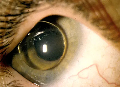
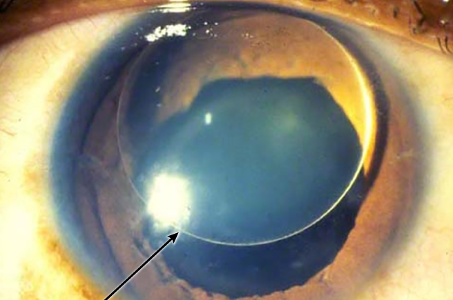
Homocystinuria is an autosomal recessive metabolic disease, most often due to a deficiency of cystathionine beta-synthase, and its most frequent eye sign is dislocation of the lens.
It resembles Marfan syndrome in body habitus, but its risk of thromboembolism and its management are different.
Recognizing the ocular signs in a child can lead to a diagnosis that changes the outcome.
What Is Homocystinuria?
Deficiency of the enzyme leads to the accumulation of homocysteine and methionine, and to a deficiency of cysteine.
The accumulation damages the zonular fibers, the vessel walls, and connective tissue, and it favors thrombosis.
The classic form is characterized by lens dislocation, a marfanoid habitus, osteoporosis, intellectual disability in some patients, and thromboembolic events.
A subset of patients respond to pyridoxine, which affects the course of the disease and the age at presentation.
Ocular Findings
- Ectopia lentis: present in most patients by adolescence, typically in an inferior or inferonasal direction
- Myopia: often high, from lens and axial changes
- Glaucoma: pupillary block by a lens in the anterior chamber or by vitreous
- Retinal detachment: more frequent than in the general population
- Cataract, optic atrophy, and corneal changes: less common
The lens subluxation often develops between ages three and ten, and it increases with time.
The zonules look fragile, and the lens may dislocate into the anterior chamber or the vitreous.
All-fit Slit-Lamp Adapter
Record and share exactly what you see at the slit lamp. One adapter fits any slit lamp or surgical microscope — and any smartphone.
From Choroida — the team behind this siteDistinguishing It From Marfan Syndrome
Ectopia lentis in Marfan syndrome is typically upward and outward, while in homocystinuria it is downward and inward.
Marfan syndrome is dominant, with aortic root dilation and joint laxity, while homocystinuria is recessive, with thrombosis, osteoporosis, and often reduced stature of the fingers and a stiff joint (see ectopia lentis in Marfan syndrome).
Weill-Marchesani syndrome has short stature and short fingers and a microspherophakic lens (see Weill-Marchesani syndrome).
A child with ectopia lentis and no clear diagnosis should have plasma amino acids and total homocysteine measured.
Diagnosis
Measure plasma total homocysteine, which is high, and plasma methionine, which is high in the classic form.
Urine tests for homocysteine are less reliable, and the diagnosis is confirmed by enzyme assay or molecular testing.
Newborn screening programs detect many cases in some countries, but later-onset forms may be missed.
Management
Systemic treatment lowers homocysteine through pyridoxine in responders, a methionine-restricted diet, betaine, folate, and vitamin B12.
Early treatment reduces the risk of thromboembolism and lens dislocation.
The ophthalmologist manages refraction and amblyopia, and follows the lens position, the pressure, and the retina.
Surgery is reserved for lens dislocation that threatens vision or that causes pupillary block, and the options are lensectomy by pars plana or limbal routes.
Surgical and Anesthetic Precautions
Patients are at risk of thrombosis during and after anesthesia, so a metabolic team should plan the perioperative care.
This includes hydration, early mobilization, and avoiding nitrous oxide, which can raise homocysteine levels.
The zonules are fragile, and the surgeon should be prepared for vitreous loss and for a lens that is in the vitreous.
Counsel families about lifelong treatment and the importance of regular follow-up.
Lens Management in Practice
Mild subluxation without visual symptoms or pupillary block can be observed, with correction of refractive error and amblyopia treatment in children.
Aphakic correction with spectacles or contact lenses is an option after lens removal, and an intraocular lens may be placed in selected patients with scleral or iris fixation.
A lens that moves into the anterior chamber can cause corneal edema and glaucoma, and it needs urgent removal.
Pupillary block can occur when the lens or the vitreous enters the pupil, and laser iridotomy, mydriatics in a supine patient, or surgery may be required.
The choice of technique depends on the patient’s age, the zonular support, and the surgeon’s experience.
Pyridoxine Responsiveness and Prognosis
Patients who respond to pyridoxine have a milder course, later lens dislocation, and fewer thromboembolic events.
Nonresponsive patients need dietary and betaine treatment, and they are at greater risk of complications.
Early detection through newborn screening and prompt treatment lead to normal intelligence and fewer eye problems in many patients.
Even treated patients need lifelong ophthalmic follow-up, because lens position and retinal status can change.
When to Think of It
Order amino acid testing in any child with spontaneous lens dislocation, particularly when the lens moves downward, and in any child with marfanoid features and developmental delay.
A history of thrombosis in a young relative, or a stroke in a child, should raise the question.
The diagnosis affects the whole family, because siblings may be affected and carriers can be identified.


Document what you see
Two smartphone imaging tools built for everyday clinic use — one for the slit lamp, one for the fundus.
From Choroida — the team behind this siteReferences
- Mudd SH, Skovby F, Levy HL, et al. The natural history of homocystinuria due to cystathionine beta-synthase deficiency. Am J Hum Genet. 1985;37:1-31.
- Burke JP, O’Keefe M, Bowell R, Naughten ER. Ocular complications in homocystinuria: early and late treated. Br J Ophthalmol. 1989;73:427-431.
- Skovby F, Gaustadnes M, Mudd SH. A revisit to the natural history of homocystinuria due to cystathionine beta-synthase deficiency. Mol Genet Metab. 2010;99:1-7.
- American Academy of Ophthalmology. Basic and Clinical Science Course, Section 6: Pediatric Ophthalmology and Strabismus. AAO; current edition.

Writes and reviews the clinical articles on Educate. Every post is checked against peer-reviewed sources before it goes live.


