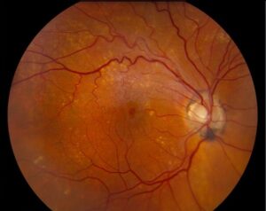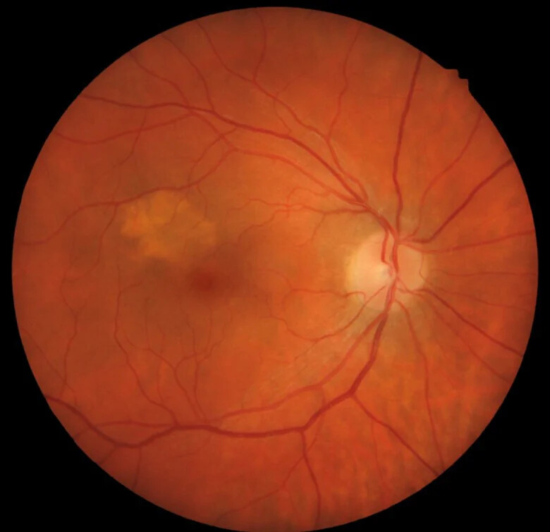
Orange pigment is a collection of lipofuscin on the surface of a melanocytic choroidal lesion.
It is a classic clinical risk factor for growth of a small pigmented lesion.
However, yellow drusen on a benign nevus can look similar at first glance.
Therefore, careful examination and autofluorescence imaging should settle the question in most eyes.
What Orange Pigment Is
Font and colleagues linked this pigment to lipofuscin, with macrophages carrying much of the material.
The deposit therefore lies at the level of the retinal pigment epithelium (RPE), over the tumor.
Lipofuscin also explains its bright autofluorescent signal (see RPE lipofuscin).
Why Orange Pigment Forms Over a Tumor
A growing tumor can disturb the overlying RPE and photoreceptors.
As a result, outer segment turnover may fail and lipofuscin may accumulate.
However, the exact sequence remains uncertain.
The pigment often accompanies subretinal fluid, which suggests a shared disturbance at the tumor surface.
Fundus Explorer Pro
Photograph the retinal findings described here with the phone already in your pocket — 22 D optics and built-in illumination in one handheld unit.
From Choroida — the team behind this siteOrange Pigment on Examination
Dilated biomicroscopy with a fundus lens usually gives the best view.
Look for orange-brown clumps or patches on the lesion surface.
In contrast, an amelanotic lesion offers little background contrast, so the pigment can escape notice.
What the Patient Reports
The pigment itself causes no symptoms.
Instead, complaints usually come from subretinal fluid or macular involvement.
Patients may then describe blur, metamorphopsia or photopsia.
Many remain asymptomatic, so routine examination often finds the lesion first.
Orange Pigment on Autofluorescence
Fundus autofluorescence gives the most useful objective evidence (see fundus autofluorescence).
True lipofuscin typically appears as dense, bright hyperautofluorescence over the lesion.
Shields and colleagues described this pattern in small choroidal melanoma.
The later Shields imaging analysis also adopted autofluorescence for documenting this risk factor.
OCT Over the Lesion
OCT adds structural context, although it cannot identify lipofuscin chemically.
Examiners may see hyperreflective material at the RPE level, often with overlying subretinal fluid.
Fluid over the lesion is an independent risk factor.
Orange Pigment as a Risk Factor
COMS Report 5 identified this sign as a predictor of growth in small melanocytic lesions.
Earlier and later Shields cohorts also supported the association.
In the 2009 analysis of 2,514 nevi, the hazard for transformation was roughly threefold.
However, these figures come from referral populations, so they may not transfer to general clinics.
Where It Fits in TFSOM
The sign supplies the “O” in the TFSOM mnemonic (see TFSOM risk factors).
Risk rises as the number of factors increases.
Therefore, one finding should always prompt a search for the others.
Thickness, fluid, symptoms, ultrasonographic hollowness and basal diameter all contribute.
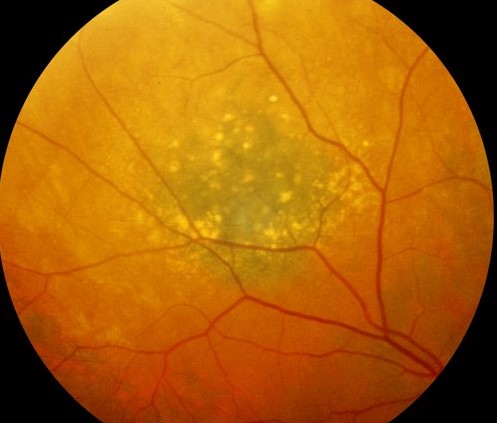
Drusen on a Nevus
Drusen often overlie chronic, stable nevi and usually suggest long duration.
Clinically, they appear as discrete, round, yellow-white dots (see drusen types).
Their presence lowers suspicion, although it never excludes melanoma by itself.
Orange Pigment Versus Drusen
Combine several findings, because no single feature settles every case.
- Color: lipofuscin looks orange-brown, while drusen look yellow-white
- Shape: lipofuscin forms irregular clumps, while drusen are usually round
- Autofluorescence: lipofuscin is intensely bright, while drusen are usually less bright
- OCT: drusen elevate the RPE from below with sub-RPE material
Workup After Finding Orange Pigment
First, take color photographs that show the lesion margins and surface.
Next, add autofluorescence and OCT through the whole lesion.
Then measure thickness and look for acoustic hollowness with B-scan ultrasonography.
Finally, record basal diameter, distance from the optic disc and any halo.
How Orange Pigment Changes Management
Confirmed lipofuscin on a suspicious lesion usually justifies ocular oncology review.
Referral should come sooner when fluid, thickness over 2 mm or documented growth coexists.
Meanwhile, a lesion with one isolated, doubtful finding may need close observation with baseline imaging.
What the Sign Cannot Do
Lipofuscin does not prove malignancy, and its absence does not prove benignity.
It also cannot estimate metastatic risk on its own.
Treatment decisions therefore rest on growth, size and location.
Counseling the Patient With Orange Pigment
Explain that the pigment is a warning sign and does not establish a cancer diagnosis.
Next, describe why imaging and specialist review help separate a nevus from small melanoma.
Patients should also report new floaters, flashes or visual distortion promptly.
Follow-Up Imaging
Repeat the same imaging modalities so that comparisons stay valid.
Intervals vary with risk, and many specialists recheck suspicious lesions within months.
In addition, keep baseline images available, because subtle change is easy to miss.
Pitfalls to Avoid
- Do not label yellow surface deposits as drusen without autofluorescence and OCT
- Never rely on one risk factor while ignoring thickness or fluid
- Do not overlook the pigment on amelanotic lesions
- Avoid comparing images from different devices without caution


Document what you see
Two smartphone imaging tools built for everyday clinic use — one for the slit lamp, one for the fundus.
From Choroida — the team behind this siteReferences
- Font RL, Zimmerman LE, Armaly MF. The nature of the orange pigment over a choroidal melanoma. Arch Ophthalmol. 1974;91:359-362.
- Collaborative Ocular Melanoma Study Group. Factors predictive of growth and treatment of small choroidal melanoma: COMS report no. 5. Arch Ophthalmol. 1997;115:1537-1544.
- Shields CL, Furuta M, Berman EL, et al. Choroidal nevus transformation into melanoma: analysis of 2514 consecutive cases. Arch Ophthalmol. 2009;127:981-987.
- Shields CL, Bianciotto C, Pirondini C, et al. Autofluorescence of choroidal melanoma in 51 cases. Br J Ophthalmol. 2008;92:617-622.
- Shields CL, Dalvin LA, Ancona-Lezama D, et al. Choroidal nevus imaging features in 3,806 cases and risk factors for transformation into melanoma in 2,355 cases. Retina. 2019;39:1840-1851.

Writes and reviews the clinical articles on Educate. Every post is checked against peer-reviewed sources before it goes live.